Figure 2.
Download original image
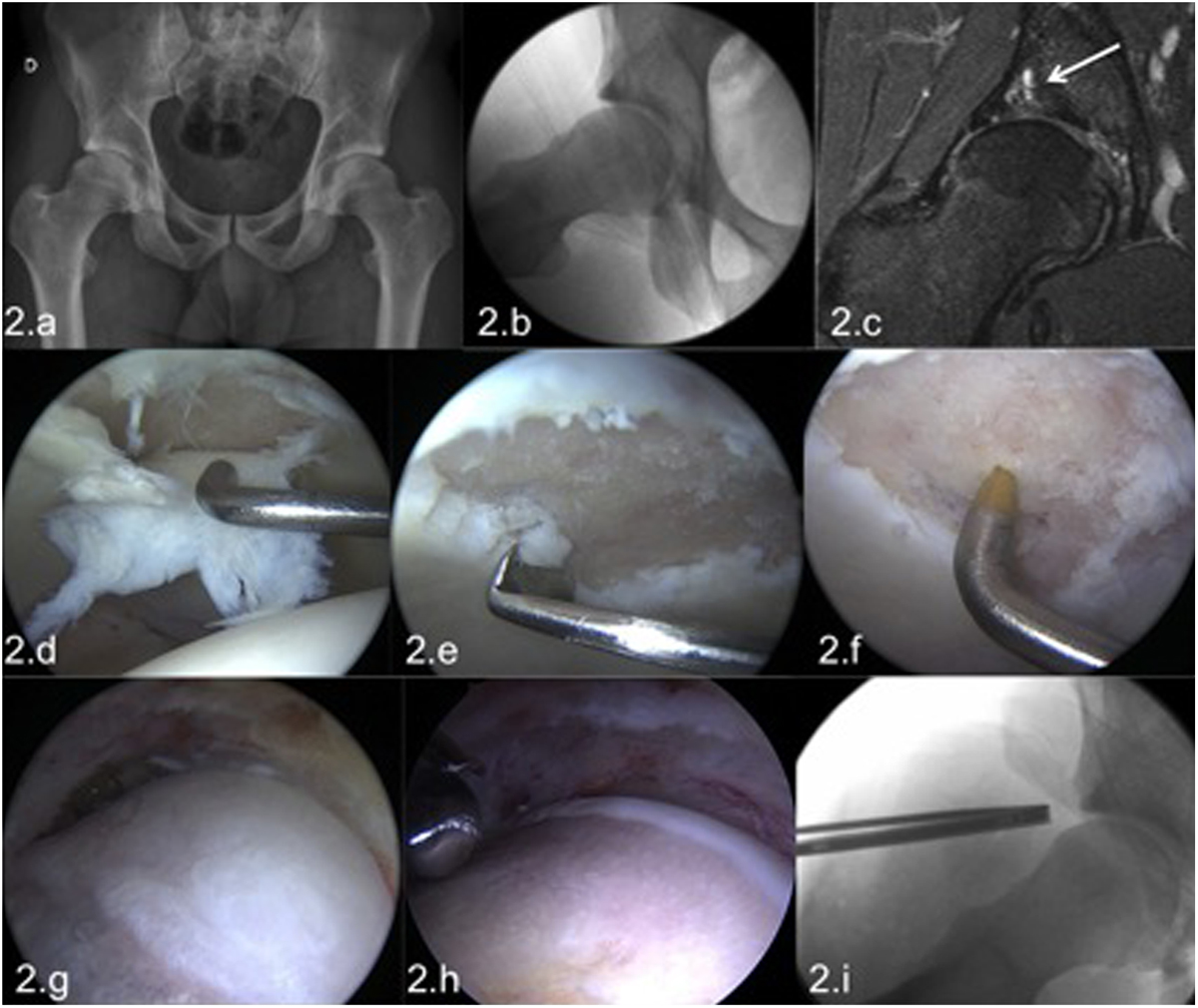
Male patient, 23 years old, medical student and amateur rugby player with sports-related pain in the right hip. The radiology demonstrated a FAI with a significant CAM deformity without significant joint space narrowing (2a, 2b). MRI showed a labral injury, a focal chondral lesion with subchondral cysts, and subchondral edema (2c). In spite of these ominous imaging signs given his young age and no other signs of osteoarthritis, hip arthroscopy was performed. At the arthroscopy an extensive full-thickness chondral lesion was found in the load-bearing surface of the acetabulum (2d); no femoral head cartilage lesions were present. The chondral lesion was treated by abrasive chondroplasty (2e) and microfractures (2f). In the peripheral compartment the extensive femoral bump (2g) was resected (2h). Intraoperative dynamic testing at the end of the femoroplasty demonstrated absence of impingement; the axial radiography demonstrated a satisfactory correction of the cam deformity (2i). Nevertheless after this arthroscopic repair, the future of this joint is uncertain with a high risk to progress to osteoarthritis in this young patient.
Current usage metrics show cumulative count of Article Views (full-text article views including HTML views, PDF and ePub downloads, according to the available data) and Abstracts Views on Vision4Press platform.
Data correspond to usage on the plateform after 2015. The current usage metrics is available 48-96 hours after online publication and is updated daily on week days.
Initial download of the metrics may take a while.