Figure 1.
Download original image
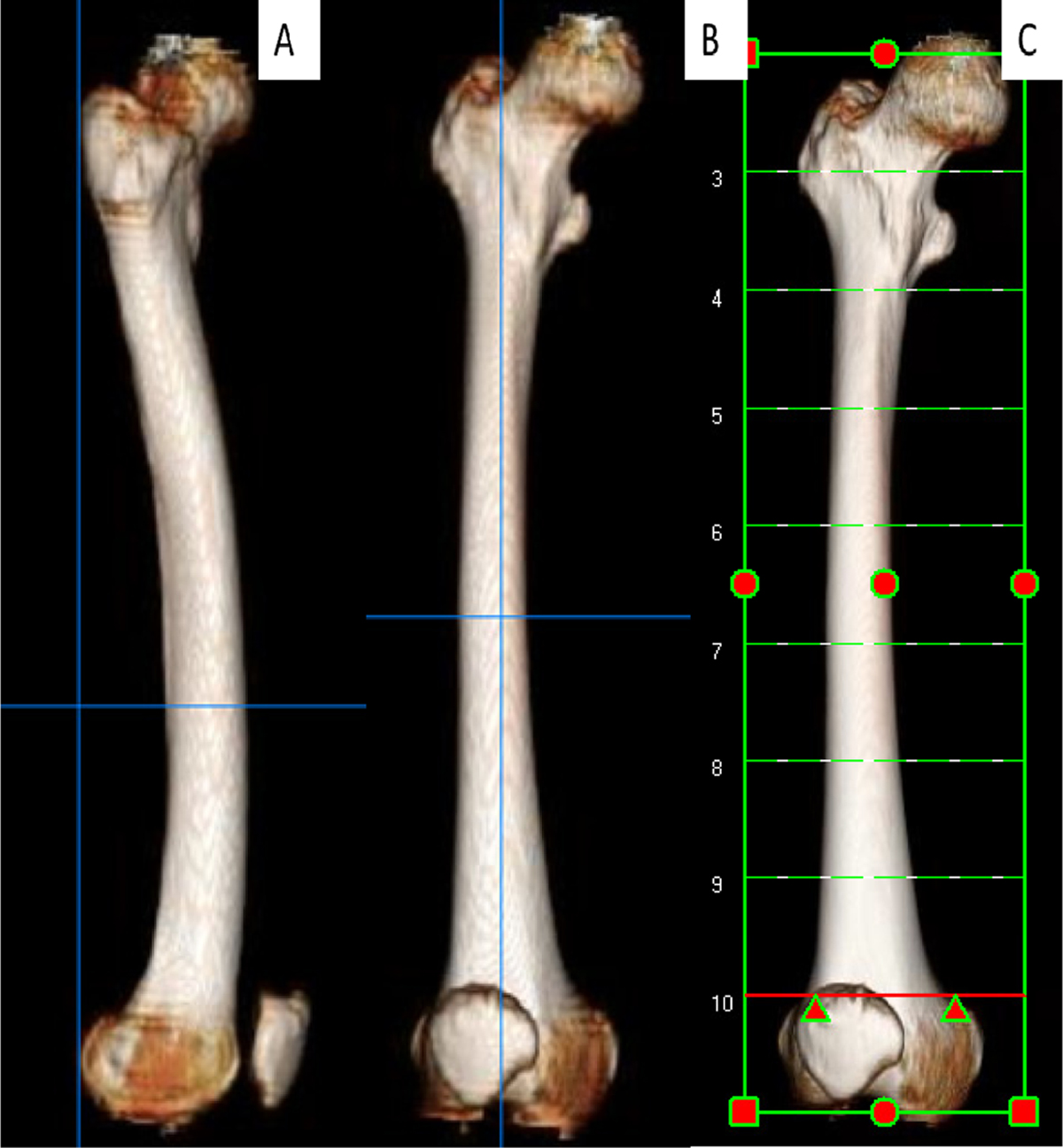
(A) Lateral view of the femur; both condyles are overlapped and a vertical line is tangential to the posterior condylar line and most posterior point of the greater trochanter, (B) anteroposterior view of the femur obtained by rotation of the lateral view 90°, (C) levels of the CT cuts starting at the widest intercondylar diameter and spaced at 5 cm intervals.
Current usage metrics show cumulative count of Article Views (full-text article views including HTML views, PDF and ePub downloads, according to the available data) and Abstracts Views on Vision4Press platform.
Data correspond to usage on the plateform after 2015. The current usage metrics is available 48-96 hours after online publication and is updated daily on week days.
Initial download of the metrics may take a while.