Figure 1.
Download original image
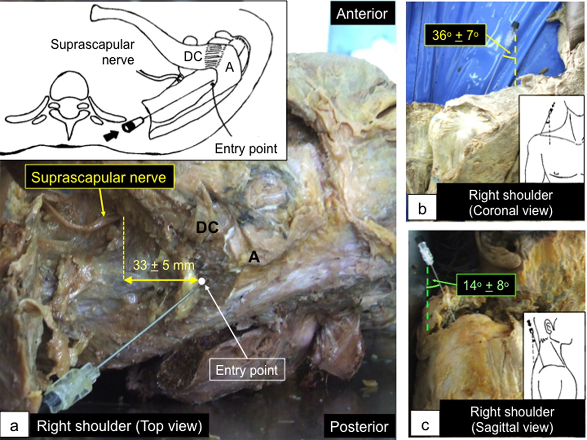
Cadaveric study of the superomedial protocol. (a) The entry point (white dot) was about 1 cm posterior to the distal clavicle and just medial to the acromion. It was about 3 cm away from the suprascapular nerve along the mediolateral axis. (b) and (c) The needle angle was 36° ± 7° lateral in the coronal plane (b) and 14° ± 8° anterior in the sagittal plane (c). A: acromion; DC: distal clavicle.
Current usage metrics show cumulative count of Article Views (full-text article views including HTML views, PDF and ePub downloads, according to the available data) and Abstracts Views on Vision4Press platform.
Data correspond to usage on the plateform after 2015. The current usage metrics is available 48-96 hours after online publication and is updated daily on week days.
Initial download of the metrics may take a while.