Figure 1.
Download original image
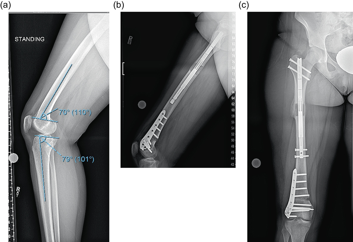
(a) This preop lateral radiograph shows a 13° flexion deformity of the distal femur. (b) Postop lateral shows a correction of the apex anterior deformity through an posterior opening wedge osteotomy stabilized with a plate and a proximal femoral lengthening with an ILN. (c) This far distal osteotomy would be difficult to control with an intramedullary implant.
Current usage metrics show cumulative count of Article Views (full-text article views including HTML views, PDF and ePub downloads, according to the available data) and Abstracts Views on Vision4Press platform.
Data correspond to usage on the plateform after 2015. The current usage metrics is available 48-96 hours after online publication and is updated daily on week days.
Initial download of the metrics may take a while.