Open Access
Figure 6.
Download original image
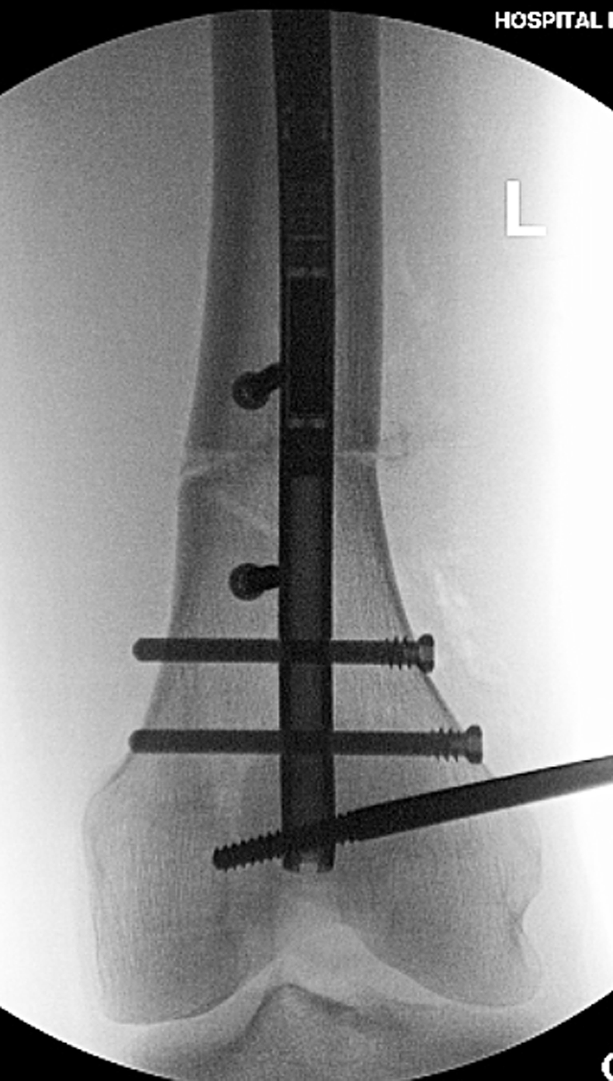
The AP fluoroscopy shot shows the distal femur after successful distal interlocking with the varus deformity corrected. The peri-osteotomy blocking screws are positioned to prevent varus deviation during lengthening. The external half pin is also seen in the field.
Current usage metrics show cumulative count of Article Views (full-text article views including HTML views, PDF and ePub downloads, according to the available data) and Abstracts Views on Vision4Press platform.
Data correspond to usage on the plateform after 2015. The current usage metrics is available 48-96 hours after online publication and is updated daily on week days.
Initial download of the metrics may take a while.