Figure 1.
Download original image
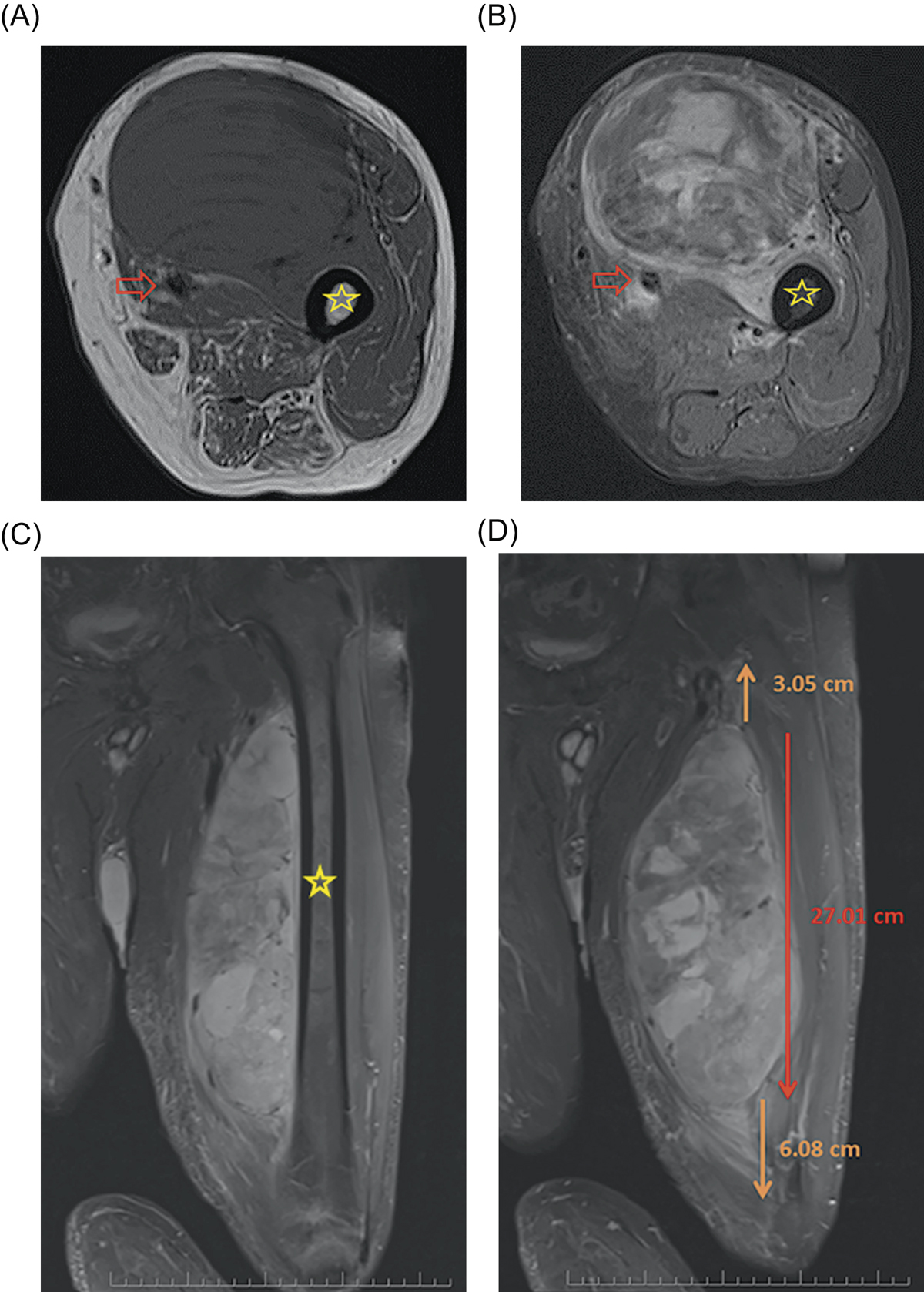
Selected T1 (A) and fat-saturated T2 (B) axial as well as fat-saturated coronal T2 (C) MRI images of a 60-year-old patient with a large, deep mass located in the anterior thigh. On the pretreatment imaging the mass was intimately associated with the femoral neurovascular bundle (arrow) as well as the periosteum of the femur (star). A biopsy was performed and showed high-grade pleomorphic rhabdomyosarcoma. The mass measured approximately 27 cm cranial/caudal however was associated with peritumoral edema which spanned nearly the entire length of the femur on coronal fat-saturated T2 (D) MRI images.
Current usage metrics show cumulative count of Article Views (full-text article views including HTML views, PDF and ePub downloads, according to the available data) and Abstracts Views on Vision4Press platform.
Data correspond to usage on the plateform after 2015. The current usage metrics is available 48-96 hours after online publication and is updated daily on week days.
Initial download of the metrics may take a while.