Figure 3.
Download original image
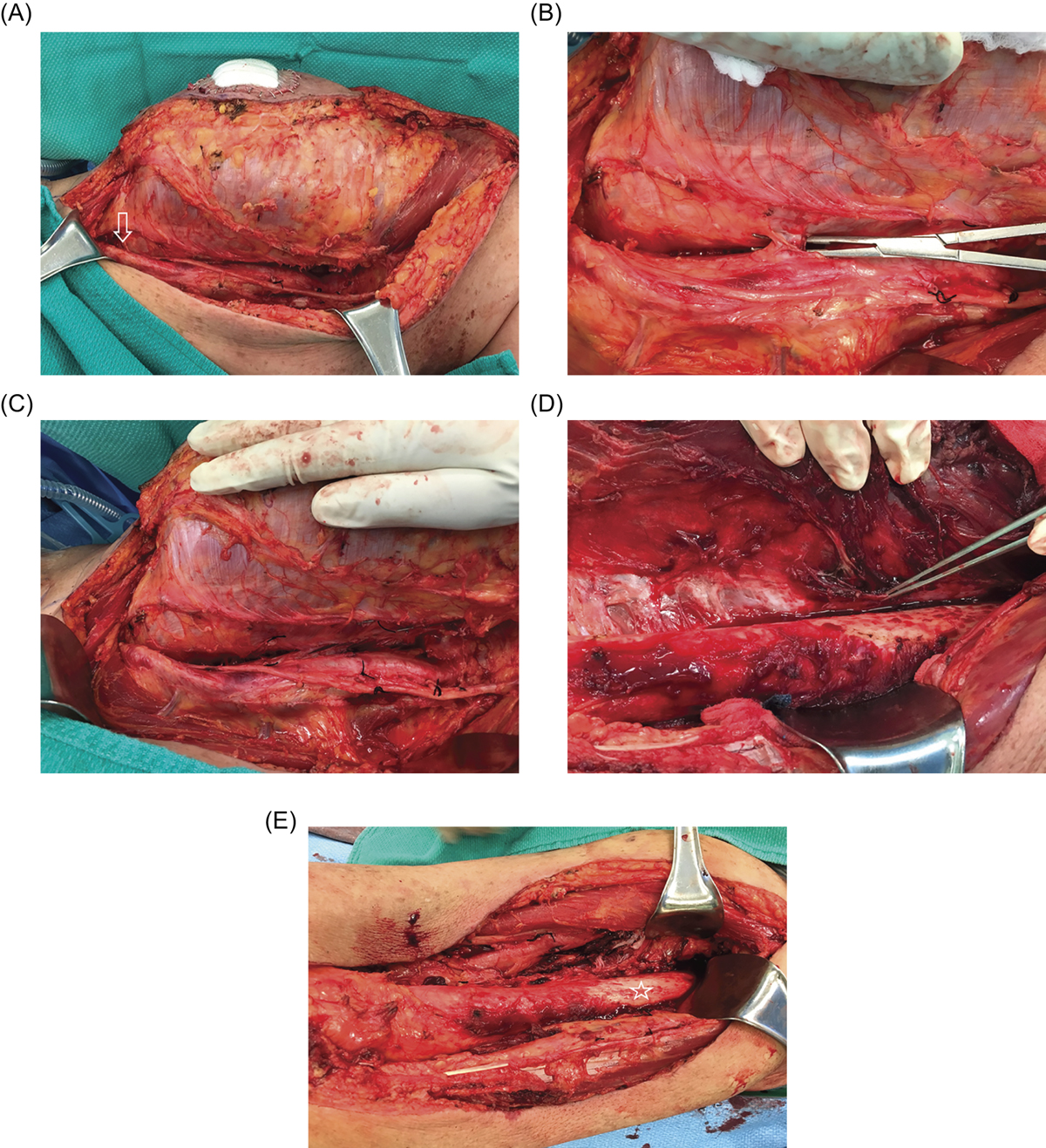
At the time of surgical excision (A), the femoral neurovascular bundle was very close to the tumor (arrow), with multiple perforating blood vessels entering the tumor (B). Due to preoperative IMRT it was safe to create a dissection plane between the tumor and the neurovascular bundle (C). The periosteum was also raised from the femur (pointer) as a margin along the tumor in the region where it was adherent to the bone (D). Although preoperative imaging showed the tumor to be very close to bone along the entire length of the femur, it was actually adherent to bone over a shorter length, so that only a small portion of the periosteum had to be removed (star) from the femoral shaft (E). The final pathological tumor resection margins were negative.
Current usage metrics show cumulative count of Article Views (full-text article views including HTML views, PDF and ePub downloads, according to the available data) and Abstracts Views on Vision4Press platform.
Data correspond to usage on the plateform after 2015. The current usage metrics is available 48-96 hours after online publication and is updated daily on week days.
Initial download of the metrics may take a while.