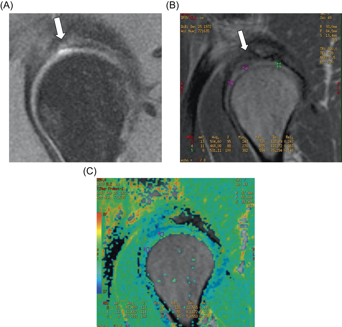
Figure 3.
Download original image
Pre- and post-operative MR of case number 1. The improvement is evaluated quantitatively (100% of filling of the lesion) and quantitatively (decrease in T2 values). (A) PDFS sagital imaging of the right hip showing the absence of cartilage in area of the anterosuperior articular surface of the acetabulum. (B, C) T2 mapping of the same hip 18 months after the surgery and the repair of the ulcerated area with improvement (decrease T2 values, showing mixed fibrocartilage and hyaline cartilage).
Current usage metrics show cumulative count of Article Views (full-text article views including HTML views, PDF and ePub downloads, according to the available data) and Abstracts Views on Vision4Press platform.
Data correspond to usage on the plateform after 2015. The current usage metrics is available 48-96 hours after online publication and is updated daily on week days.
Initial download of the metrics may take a while.