Figure 3
Download original image
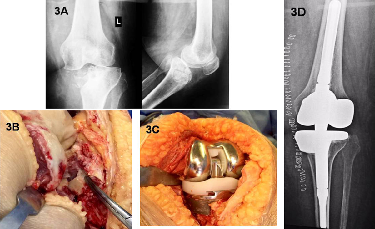
(A) Preoperative X-rays for a depressed fracture of the medial tibial plateau. Metaphyseal collapse and knee subluxation can be observed. (B) Intraoperative photo shows unreconstructable comminution of the articular surface. (C) The bone defect was reconstructed using a medial 20 mm metal step attached to the tibial tray. (D) Postoperative X-ray showing the Rotating Hinge implant and metal augment to the proximal tibia.
Current usage metrics show cumulative count of Article Views (full-text article views including HTML views, PDF and ePub downloads, according to the available data) and Abstracts Views on Vision4Press platform.
Data correspond to usage on the plateform after 2015. The current usage metrics is available 48-96 hours after online publication and is updated daily on week days.
Initial download of the metrics may take a while.