Figure 1.
Download original image
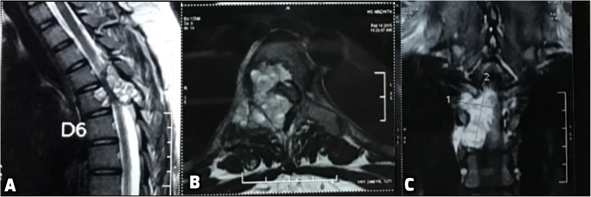
First Pre-operative MRI (A) revealing an intensely enhancing cystic cum haemorrhagic septate expansile bony lesion (5.7 × 3.1 cm) involving the posterior part of D5 vertebral body, right lamina, right pedicle and transverse process with epidural extension (D4–D5) causing marked spinal cord compression and underlying cord oedema. (B) Axial T2 MRI scan depicts the encroachment of the right half of body and posterior elements with compression of the spinal cord. (C) Coronal image through the spinal canal showing intense enhancement on T2 image with fluid levels.
Current usage metrics show cumulative count of Article Views (full-text article views including HTML views, PDF and ePub downloads, according to the available data) and Abstracts Views on Vision4Press platform.
Data correspond to usage on the plateform after 2015. The current usage metrics is available 48-96 hours after online publication and is updated daily on week days.
Initial download of the metrics may take a while.