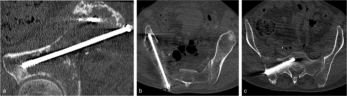
Figure 2
Download original image
(a) Oblique sagittal reformatted view of the pelvis CT scan (bone window, slice thickness 1 mm) acquired after osteosynthesis (first step of the procedure). The first screw was placed into the iliac bone from the posterior iliac crest to the anterior inferior iliac spine along the arcuate line. The cross section of the proximal part of the second screw inserted into the sacroiliac joint is visible (small arrow). (b) Oblique axial reformatted view of the pelvis CT scan after iliac screw fixation. (c) Oblique axial reformatted view of the pelvis CT scan after sacral screw fixation. This second screw was inserted into the sacroiliac joint to the sacral wing.
Current usage metrics show cumulative count of Article Views (full-text article views including HTML views, PDF and ePub downloads, according to the available data) and Abstracts Views on Vision4Press platform.
Data correspond to usage on the plateform after 2015. The current usage metrics is available 48-96 hours after online publication and is updated daily on week days.
Initial download of the metrics may take a while.