Figure 4
Download original image
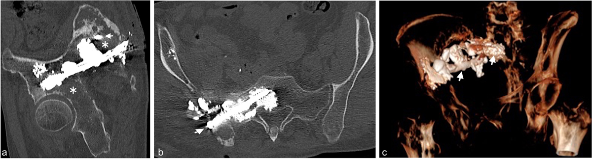
(a) Oblique sagittal reformatted view of post-procedure CT (bone window, slice thickness 1 mm) of the pelvic bones after inserting screws and first step of cementoplasty. The cross section of the proximal part of the sacral screw is visible (small arrow). After this procedure, two large osteolytic areas compromising the whole stability of the osteosynthesis remained, one in the right acetabulum and the other in the right posterior iliac wing with no cement continuity between the two screws at that step (asterisk). (b) Oblique coronal reformatted view of post-procedure CT (bone window, slice thickness 1 mm) of the pelvic bones after inserting screws and first step of cementoplasty shows the sacral screw (small arrow) anchored into the bone with cementoplasty and a slight extravasation of bone cement in soft-tissue. (c) 3D oblique anterior reformatted view of post-procedure CT shows the whole osteosynthesis with the two screws (small arrows) surrounded by cement.
Current usage metrics show cumulative count of Article Views (full-text article views including HTML views, PDF and ePub downloads, according to the available data) and Abstracts Views on Vision4Press platform.
Data correspond to usage on the plateform after 2015. The current usage metrics is available 48-96 hours after online publication and is updated daily on week days.
Initial download of the metrics may take a while.