Figure 4
Download original image
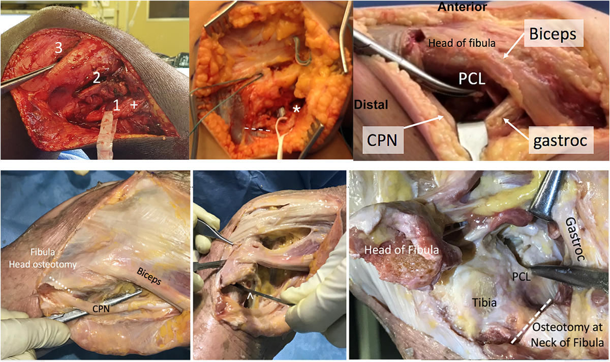
Lateral approach to careful dissection and retraction of common peroneal nerve (CPN). Top left: release of the nerve through window “2”, in which the nerve has (+) a drop-down appearance with appropriate release below window “1” and the forceps are placed into window “3”. Top middle: release of peroneal nerve (*) in a chronic condition with extensive scarring and release to peroneus longus fascia (white line). Top right: lateral Lobenhoffer approach without fibula head osteotomy in a cadaver, in which protecting the CPN can be achieved through access distal to the biceps tendon, retracting the gastrocnemius (“gastroc”) tendon posterior. The tibial insertion of the posterior cruciate ligament (“PCL”) can be reached posterior to the head of the fibula. Bottom left: lateral Lobenhoffer approach to the posterior part of the tibia in a cadaver. Initially, the CPN is identified and protected. Bottom middle and right: An osteotomy at the neck of the fibula (^) increases access to the PCL when the gastrocnemius and popliteus muscles are retracted posteriorly.
Current usage metrics show cumulative count of Article Views (full-text article views including HTML views, PDF and ePub downloads, according to the available data) and Abstracts Views on Vision4Press platform.
Data correspond to usage on the plateform after 2015. The current usage metrics is available 48-96 hours after online publication and is updated daily on week days.
Initial download of the metrics may take a while.