Figure 4
Download original image
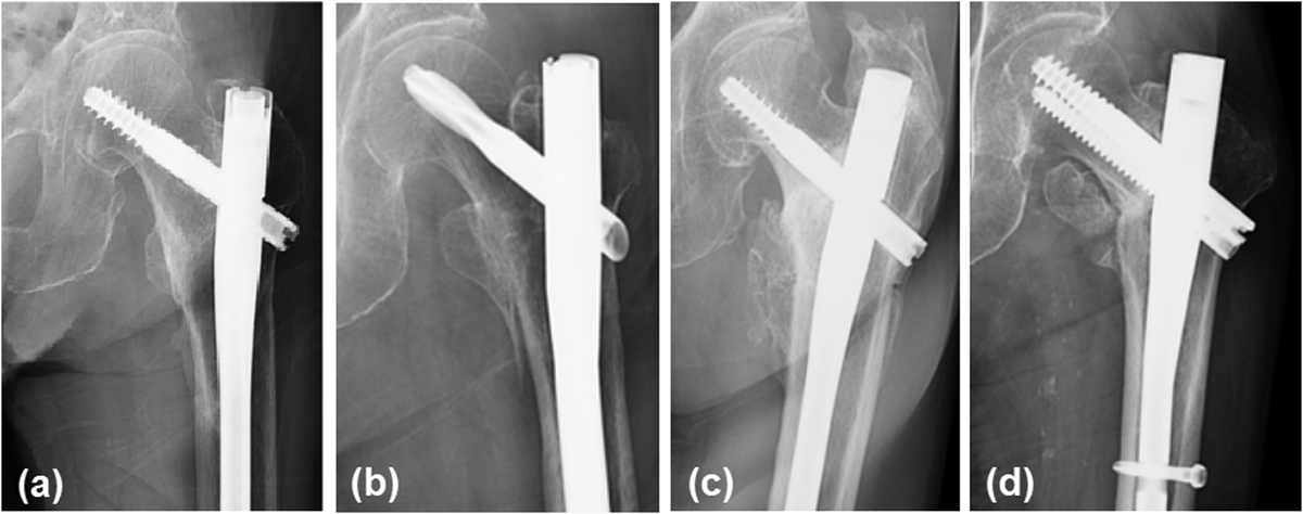
These radiographs show the deformity degree. (a) The radiograph presented mild deformity but showed complete bone union. Slight ossification was observed around the tape. (b) The radiograph presented moderate deformity but also showed complete bone union with 5–10 mm displacement of the lesser trochanter fragment. (c) The radiograph presented severe deformity with the displacement of lesser trochanter fragment to >1 cm from the base. (d) The radiograph showed ununited fragment. The displaced fragment shifted from the original position and was observed in the anterior proximal aspect of the femur. This may be affected by the iliopsoas muscle force.
Current usage metrics show cumulative count of Article Views (full-text article views including HTML views, PDF and ePub downloads, according to the available data) and Abstracts Views on Vision4Press platform.
Data correspond to usage on the plateform after 2015. The current usage metrics is available 48-96 hours after online publication and is updated daily on week days.
Initial download of the metrics may take a while.