Figure 1
Download original image
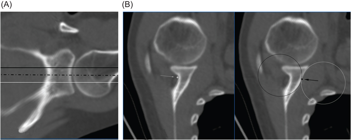
(A) CT imaging showing the position of the three axial cross-sections used to determine the GvOC ROI landmarks; black line; showing the axial cross-section at the level of the superior third-middle third junction; white line; showing the junction between the middle-third and inferior third; dotted line; showing the equatorial level of the glenoid. (B) CT imaging showing axial cross-sections of a glenoid specimen analysed with the GvOC ROI landmarks demonstrated. The left image; posterior ROI point – white arrow, white dot. The right image; The anterior slope of the glenoid is slightly curved with an anterior concavity and therefore fits with a sphere as demonstrated by the white sphere; and the onset of the subscapularis fossa also has a curved shape, with a posterior concavity, that also fits with a sphere as demonstrated by the black sphere. The anterior ROI point – black point, black arrow – is placed at the cross-section between both spheres, that also represents the change of curvature of the anterior aspect of the glenoid vault.
Current usage metrics show cumulative count of Article Views (full-text article views including HTML views, PDF and ePub downloads, according to the available data) and Abstracts Views on Vision4Press platform.
Data correspond to usage on the plateform after 2015. The current usage metrics is available 48-96 hours after online publication and is updated daily on week days.
Initial download of the metrics may take a while.