Figure 2
Download original image
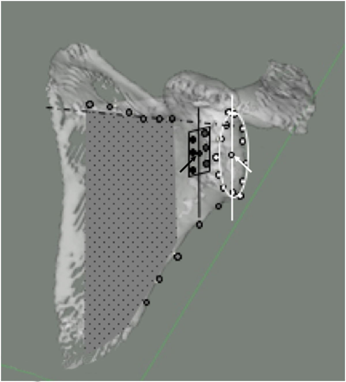
3D reconstruction of a scapula specimen showing the various radiological reference planes. GR plane (represented by the oval shape with white border) of points placed at the edge of the articular surface (White ROI points with black contour), GR centre (white arrow), and GR superior-inferior axis (white line). B plane (Grey doted area) formed by points (Grey ROI points with black contour) placed on the spine root pf the scapula (dotted line) and on the lateral border of the scapula. New GvOC plane (represented by the black rectangle) formed by points (Black ROI points) on the six described GvOC ROI landmarks, with best fit centre (Black arrow) and superior-inferior direction (Black line).
Current usage metrics show cumulative count of Article Views (full-text article views including HTML views, PDF and ePub downloads, according to the available data) and Abstracts Views on Vision4Press platform.
Data correspond to usage on the plateform after 2015. The current usage metrics is available 48-96 hours after online publication and is updated daily on week days.
Initial download of the metrics may take a while.