Figure 9
Download original image
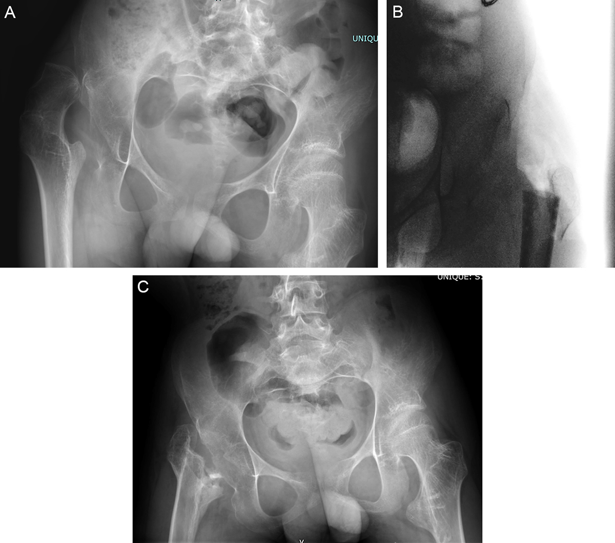
(A) Preoperative radiograph of 18-year-old male, GMFCS III/IV with right hip pain and difficulty with weight bearing and activities of daily living. Note the asphericity of the femoral head and skeletal maturity indicating that remodeling of the femoral head here was not going to be substantive. (B) Intraoperative radiograph demonstrating the relationship between the trochanteric fragment and the femoral shaft. These pieces of bone are transfixed with #2 Fibrewire sutures. (C) Final 1-year radiograph of hip salvage intervention. This young man has returned to weight bearing without pain and his overall function has improved significantly. Note the appropriate station of the proximal femur without significant migration and pelvic support configuration.
Current usage metrics show cumulative count of Article Views (full-text article views including HTML views, PDF and ePub downloads, according to the available data) and Abstracts Views on Vision4Press platform.
Data correspond to usage on the plateform after 2015. The current usage metrics is available 48-96 hours after online publication and is updated daily on week days.
Initial download of the metrics may take a while.