Figure 1
Download original image
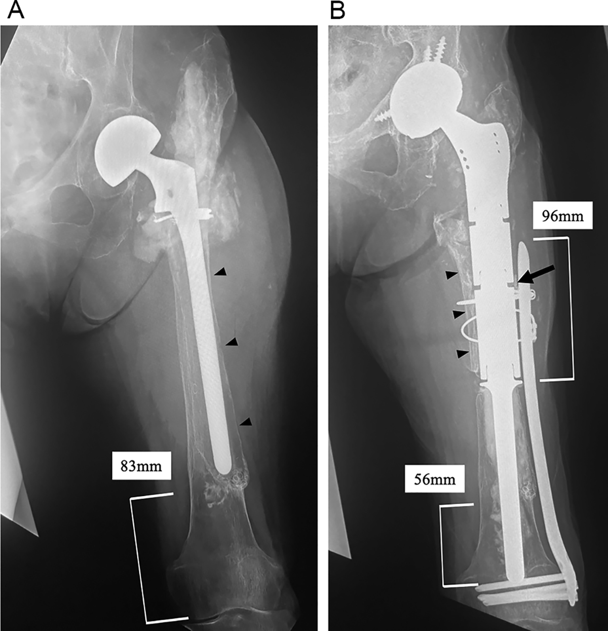
(A) A preoperative radiograph of the femur demonstrating stem loosening and massive bone loss from the proximal to the distal end of the diaphyseal femur (Paprosky type IV) with acetabular osteolysis on the left side. The radiolucent line surrounding the stem and thin cortical bone of the femur are observed (black arrowheads). Additionally, severe stem migration as a consequence of the loosening is identified. The intact distal femoral bone remains only approximately 83 mm from the distal end of the femur. In addition, its bone quality is osteoporotic. (B) A postoperative radiograph of the femur 5 years after surgery demonstrates no signs of a radiolucent line, implant-related issues, or bone resorption. The PFR stem is inserted 56 mm into the intact distal femur (approximately 45% of the PFR stem length). A locking plate is placed on the femur, overlapping the PFR 96 mm proximally from the bone-cement-prosthesis junction. The black arrowheads indicate the longitudinally opened bone fragment of the proximal diaphyseal femur. The black arrow indicates the free cortical bone fragment between the PFR and locking plate.
Current usage metrics show cumulative count of Article Views (full-text article views including HTML views, PDF and ePub downloads, according to the available data) and Abstracts Views on Vision4Press platform.
Data correspond to usage on the plateform after 2015. The current usage metrics is available 48-96 hours after online publication and is updated daily on week days.
Initial download of the metrics may take a while.