Figure 5
Download original image
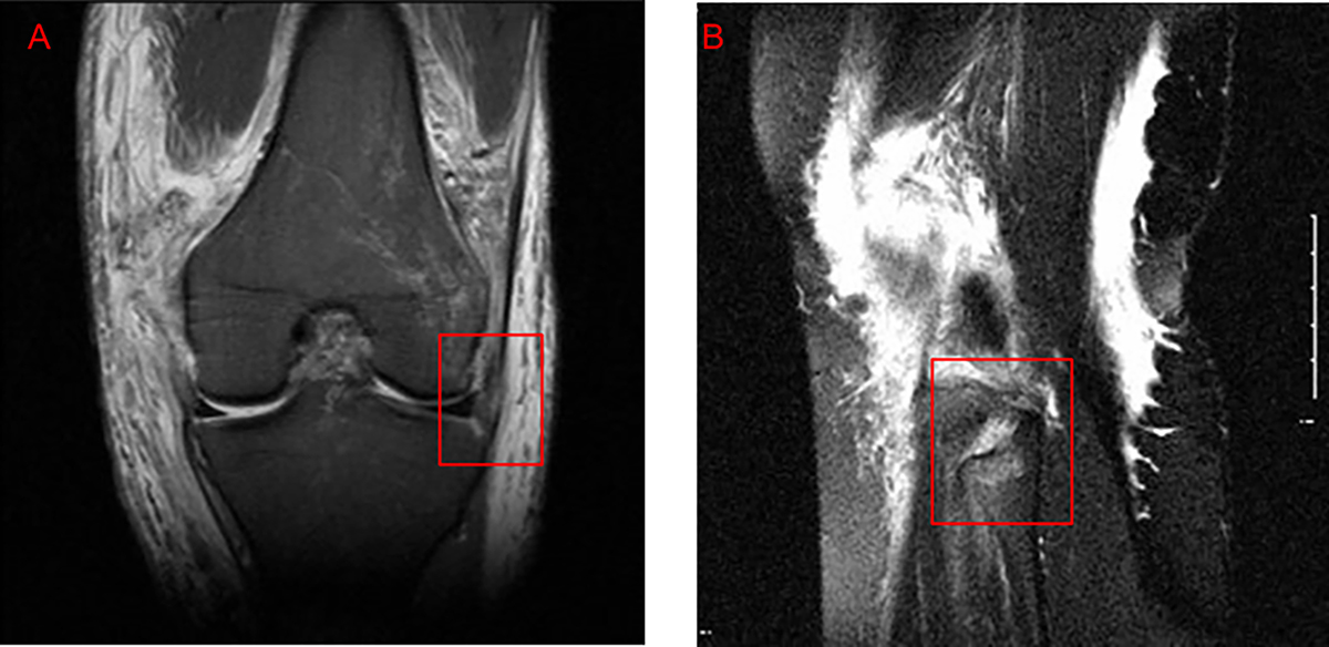
Examples of posterolateral corner (PLC) injuries that were misinterpreted as intact on pre-operative MRI. (A) Coronal fat-suppressed proton-density image shows a full-thickness tear of the iliotibial band (box), reported as intact in the original radiology report. (B) Sagittal proton-density image from a different case that demonstrates disinsertion of the biceps femoris tendon with bone marrow edema of the fibular head (box), also misreported as intact.
Current usage metrics show cumulative count of Article Views (full-text article views including HTML views, PDF and ePub downloads, according to the available data) and Abstracts Views on Vision4Press platform.
Data correspond to usage on the plateform after 2015. The current usage metrics is available 48-96 hours after online publication and is updated daily on week days.
Initial download of the metrics may take a while.