Figure 2
Download original image
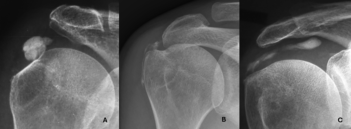
Representative imaging features of Type I and Type II calcifications. A. Type I – In situ calcification confined to the tendon, without signs of migration. Note the cortical sclerosis of the greater tuberosity adjacent to the deposit. B. Type II-a – Extension into the subacromial-subdeltoid bursa. C. Type II-b – Medial progression along the supraspinatus tendon, creating a characteristic “comet-tail” appearance.
Current usage metrics show cumulative count of Article Views (full-text article views including HTML views, PDF and ePub downloads, according to the available data) and Abstracts Views on Vision4Press platform.
Data correspond to usage on the plateform after 2015. The current usage metrics is available 48-96 hours after online publication and is updated daily on week days.
Initial download of the metrics may take a while.