Figure 3
Download original image
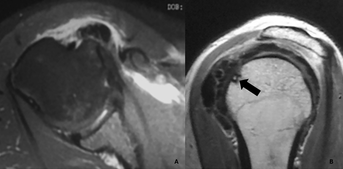
Magnetic resonance imaging of a Type III-a calcification in the subscapularis tendon. A. Axial view showing a calcific deposit in direct contact with the cortex of the lesser tuberosity, associated with marked subcoracoid bursitis. B. Oblique sagittal view demonstrating focal cortical erosion at the site of contact (black arrow), without evidence of intraosseous extension.
Current usage metrics show cumulative count of Article Views (full-text article views including HTML views, PDF and ePub downloads, according to the available data) and Abstracts Views on Vision4Press platform.
Data correspond to usage on the plateform after 2015. The current usage metrics is available 48-96 hours after online publication and is updated daily on week days.
Initial download of the metrics may take a while.