Figure 4
Download original image
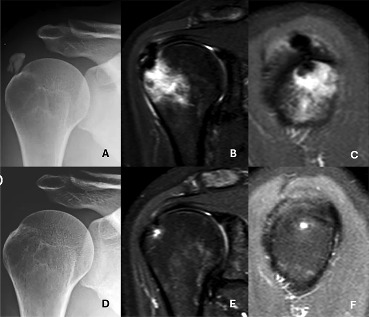
Advanced intraosseous migration (Type III-b) of a supraspinatus calcification with spontaneous resolution at follow-up. A. Initial anteroposterior (AP) radiograph showing the calcific deposit. B. Coronal MRI revealing intraosseous extension of the deposit with associated bone marrow edema. C. Oblique sagittal MRI demonstrating the “hourglass sign,” indicating tendon-to-bone continuity. D. Follow-up AP radiograph two years later showing complete disappearance of the calcification. E. Coronal MRI confirming resolution of the intraosseous deposit. F. Oblique sagittal MRI showing complete resolution of both the calcification and bone marrow edema.
Current usage metrics show cumulative count of Article Views (full-text article views including HTML views, PDF and ePub downloads, according to the available data) and Abstracts Views on Vision4Press platform.
Data correspond to usage on the plateform after 2015. The current usage metrics is available 48-96 hours after online publication and is updated daily on week days.
Initial download of the metrics may take a while.