Figure 3
Download original image
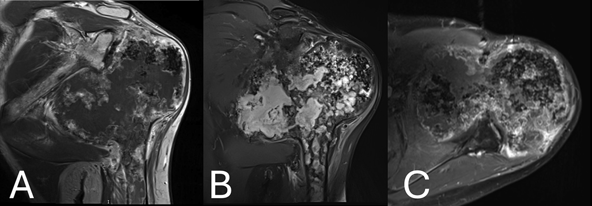
(A) Magnetic resonance imaging showing the exuberant medial compartment, denoted as a large lobulated area, extending to the cortex, with low signal intensity on T1W. (B) Magnetic resonance imaging showing High signal intensity on fat-suppressed T2 W images of the lobulated area on the MRI. Flocculent stippled areas of a signal void, representing cartilaginous calcifications, were more prominent in the lateral compartment. Geographic areas of marrow signal, which are high on T1W and low on fat-suppressed T2W, were entrapped within the cartilaginous tissue of the medial compartment, whereas bone marrow signal was seen at the periphery of the lateral compartment. Cortical disruption was seen at the posterior aspect of the epiphysis corresponding to the osteolytic areas shown on CT. (C) Magnetic resonance imaging post gadolinium administration showing mild peripheral inhomogeneous enhancement. Tissue enhancement was more diffuse or nodular and somewhat more intense at the posterior aspect adjacent to the osteolytic area. Lobules of cartilaginous signal intensity were also seen in the middle and lateral components of the epiphysis, extending to the metaphysis.
Current usage metrics show cumulative count of Article Views (full-text article views including HTML views, PDF and ePub downloads, according to the available data) and Abstracts Views on Vision4Press platform.
Data correspond to usage on the plateform after 2015. The current usage metrics is available 48-96 hours after online publication and is updated daily on week days.
Initial download of the metrics may take a while.