Figure 6
Download original image
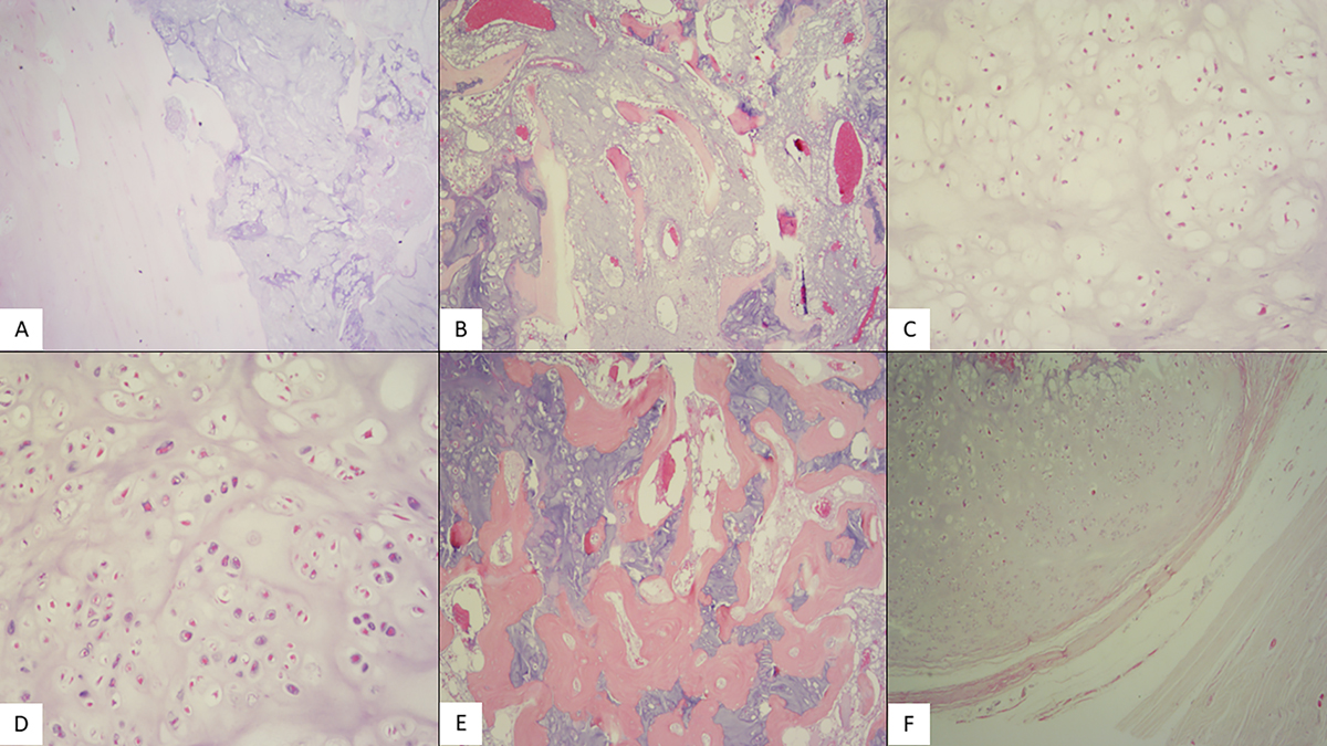
(A) Hematoxylin-eosin stain (X20): Low power shows compact bone infiltration by a chondroid neoplasm. (B) Hematoxylin-eosin stain (X40): Hyaline cartilage lobules expand between spongy bone trabeculae. (C) Hematoxylin-eosin stain (X100): The tumor is composed of hyaline cartilage lobules with mild cellular atypia (chondrosarcoma grade 1) and (D) Hematoxylin-eosin stain (X100): Medium cellular atypia (chondrosarcoma grade 2). Some neoplastic cells are binucleated. Mitoses are rare. (E) hematoxylin-eosin stain (X40): Evident infiltration of the haversian system. (F) Hematoxylin-eosin stain (X40): Extension to periosteal soft tissue.
Current usage metrics show cumulative count of Article Views (full-text article views including HTML views, PDF and ePub downloads, according to the available data) and Abstracts Views on Vision4Press platform.
Data correspond to usage on the plateform after 2015. The current usage metrics is available 48-96 hours after online publication and is updated daily on week days.
Initial download of the metrics may take a while.