Figure 1
Download original image
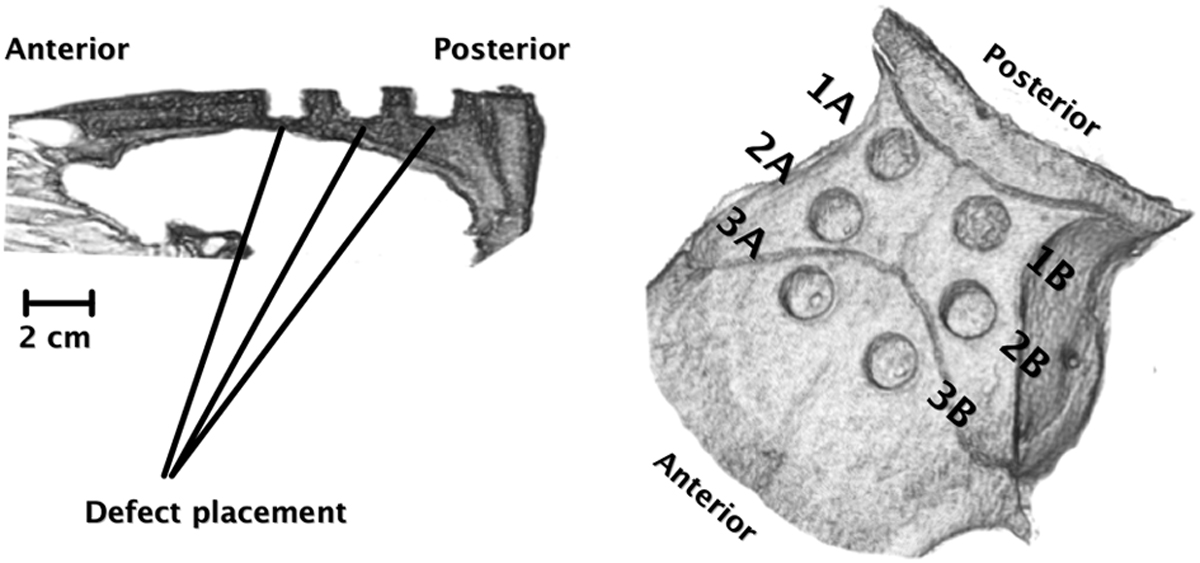
CT reconstructions one week after surgery (0.6 mm thick, non-overlapping sections, 60 mA, 120 kV) acquired using a SOMATOM Definition CT scanner (Siemens AG Medical Solutions, Erlangen, Germany) and visualized using OsiriX v.5.7.1 64-bit (Bernex, Switzerland). The left reconstruction illustrates a sagittal view of the position and depth of the drill holes in relation to the cranial cavity. Note that the calvaria thickness decreases toward the anterior part. On the right, the three sub-studies, 1A/1B, 2A/2B, and 3A/3B, are illustrated in relation to the cranial sutures.
Current usage metrics show cumulative count of Article Views (full-text article views including HTML views, PDF and ePub downloads, according to the available data) and Abstracts Views on Vision4Press platform.
Data correspond to usage on the plateform after 2015. The current usage metrics is available 48-96 hours after online publication and is updated daily on week days.
Initial download of the metrics may take a while.