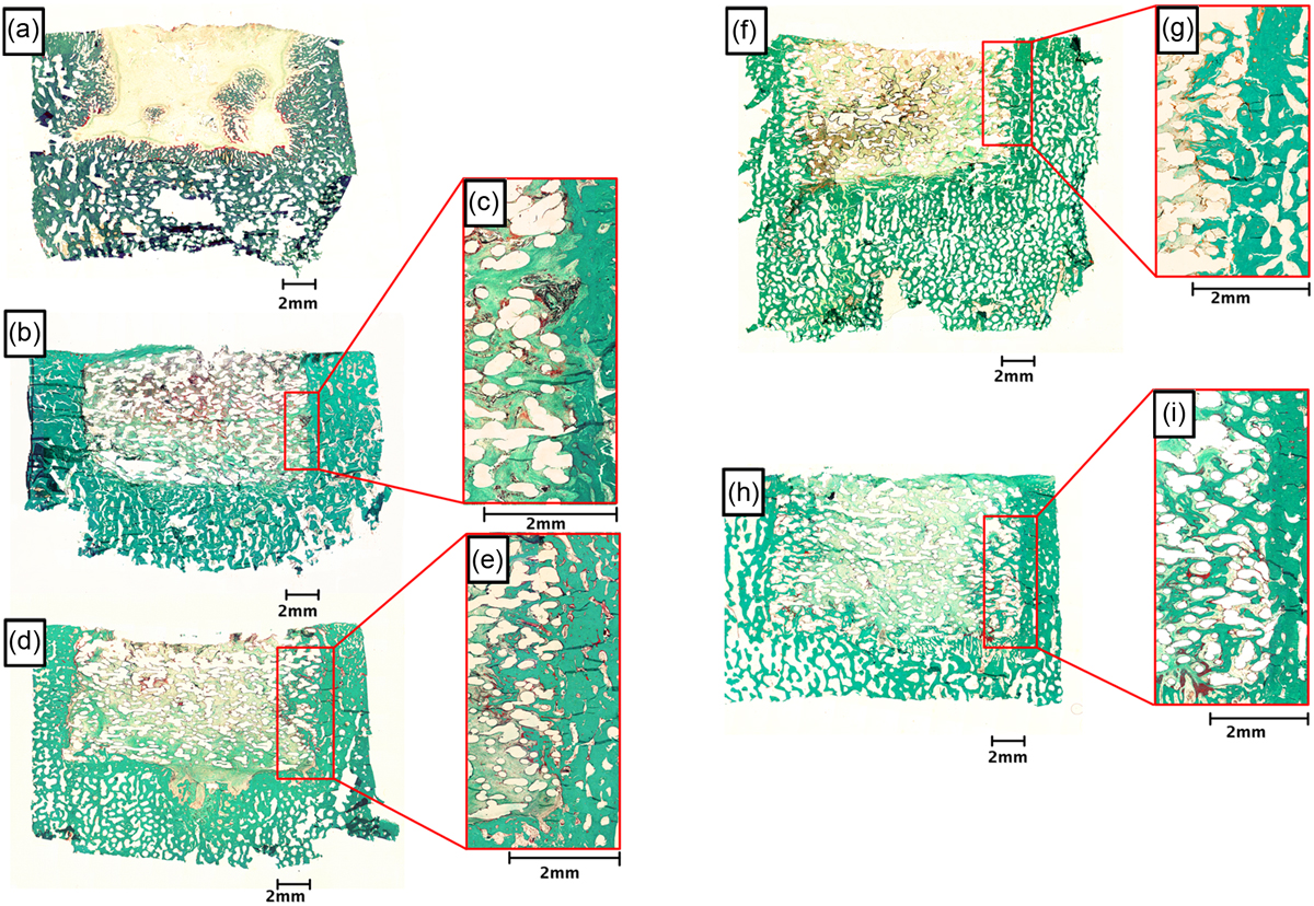
Figure 5.
Download original image
Representative images of histological sections stained with Goldner’s Trichrome. The histological sections depict: (a) empty defect or defects treated with (b) PCL scaffold; (d) HT-CPL scaffold; (f) BMSCs on HT-PCL scaffolds; and (h) DPSCs on HT-PCL scaffolds. Superimposed magnifications of the transition area between defect and existing bone are presented for each of the scaffold-containing groups. The magnifications are: (c) PCL scaffold; (e) HT-CPL scaffold; (g) BMSCs on HT-PCL scaffolds, and (i) DPSCs on HT-PCL scaffolds. Translucent areas within the defects treated with a PCL scaffold represent areas where PCL was present prior to embedding. Scale bars are presented at each image.
Current usage metrics show cumulative count of Article Views (full-text article views including HTML views, PDF and ePub downloads, according to the available data) and Abstracts Views on Vision4Press platform.
Data correspond to usage on the plateform after 2015. The current usage metrics is available 48-96 hours after online publication and is updated daily on week days.
Initial download of the metrics may take a while.