Figure 4.
Download original image
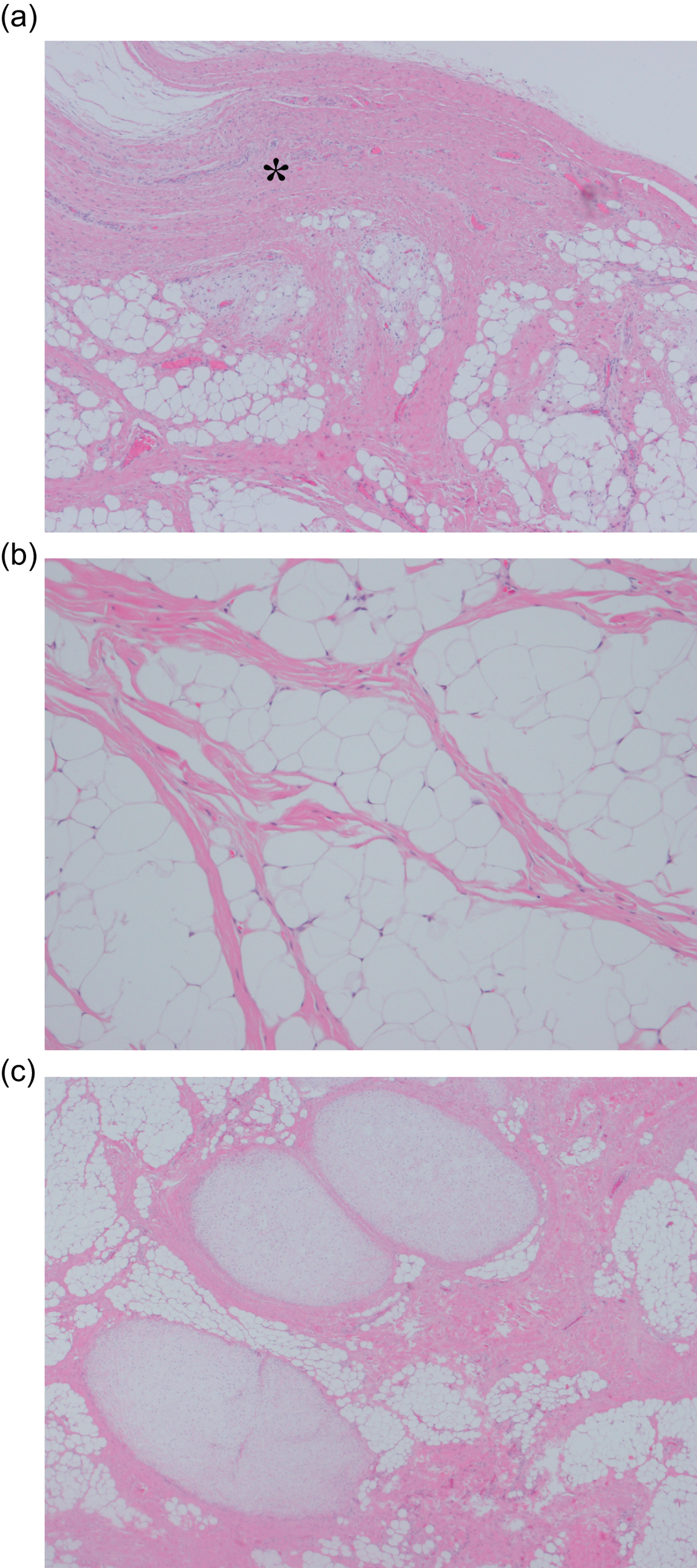
Histopathological examination of excised tumor. (a) Synovial tissue was detected in the margin of this tumor (*). In the subsynovia, there was a thick fibrous septum and diffuse proliferation of fat tissue (Hematoxylin and eosin, × 20). (b) Diffuse proliferation of fat tissue was also detected in the tumor and atypical lipoblasts were not seen (Hematoxylin and eosin, × 50). (c) A few chondral nodules and a thick fibrous septa were found in the fat tissue (Hematoxylin and eosin, × 10).
Current usage metrics show cumulative count of Article Views (full-text article views including HTML views, PDF and ePub downloads, according to the available data) and Abstracts Views on Vision4Press platform.
Data correspond to usage on the plateform after 2015. The current usage metrics is available 48-96 hours after online publication and is updated daily on week days.
Initial download of the metrics may take a while.