Figure 2.
Download original image
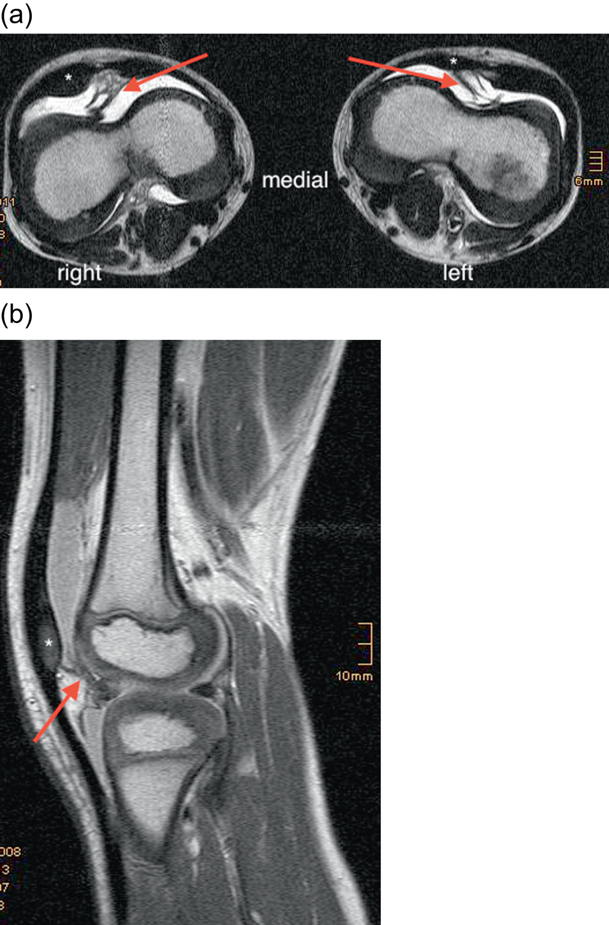
(a) Preoperative axial MRI of both knees in a six-year-old boy shows a deep femoral trochlea with a septum (red arrow), dividing the knee joint in a medial and lateral half and positioning the patella on the lateral side of the septum; patella ossification center (*). (b) Sagittal MRI through the lateral femoral condyle articulating with the patella ossification center (*); septum (red arrow).
Current usage metrics show cumulative count of Article Views (full-text article views including HTML views, PDF and ePub downloads, according to the available data) and Abstracts Views on Vision4Press platform.
Data correspond to usage on the plateform after 2015. The current usage metrics is available 48-96 hours after online publication and is updated daily on week days.
Initial download of the metrics may take a while.