Figure 2.
Download original image
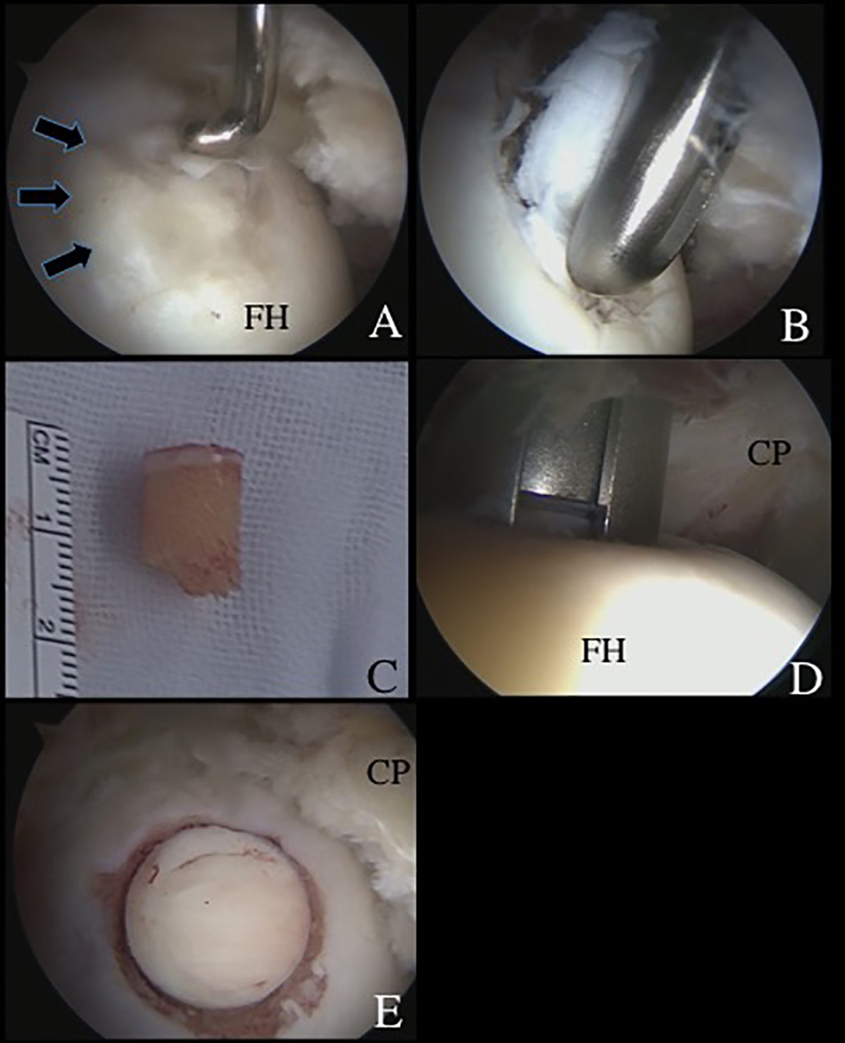
Surgical findings and technique. (A) An osteochondritis dissecans (OCD) lesion (arrows), classified as International Cartilage Repair Society grade III, was observed at the anterosuperior femoral head, with the arthroscope viewing from the anterolateral portal (ALP). A probe through the proximal midanterior portal (PMAP) was used to evaluate the OCD lesion. (B) The degenerative OCD lesion was resected under direct visualization from the ALP. (C) A cylindrical autologous osteochondral graft (8.5 mm in diameter) was harvested arthroscopically from the ipsilateral knee joint. (D) A drill guide was introduced through the PMAP, viewing from the ALP. The subchondral bone was drilled to a depth of 14 mm. The dilator was inserted into the drill guide and tapped to the desired depth. (E) The autologous osteochondral graft was tamped into the lesion until the articular surface was flush with the host joint surface. (CP: capsule; FH: femoral head.)
Current usage metrics show cumulative count of Article Views (full-text article views including HTML views, PDF and ePub downloads, according to the available data) and Abstracts Views on Vision4Press platform.
Data correspond to usage on the plateform after 2015. The current usage metrics is available 48-96 hours after online publication and is updated daily on week days.
Initial download of the metrics may take a while.