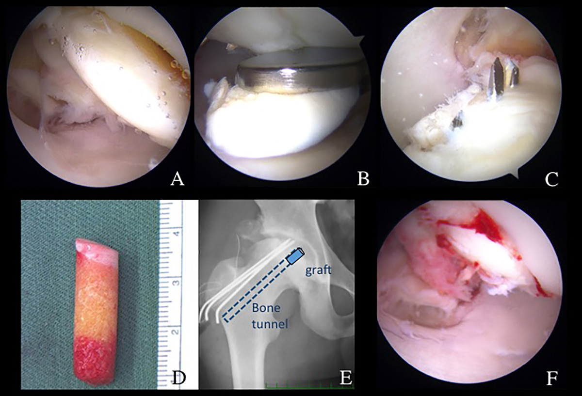
Figure 6.
Download original image
(A) Arthroscopic finding from ALP showing OCD lesion separated from the femoral head. (B) Viewing from MAP, OCD fragment was removed by forceps through ALP. (C) Viewing from PMAP, OCD was fixed by three 2 mm K-wires through CROSSTRAC guide. (D) Cylindrical osteochondral autograft harvested from femur. (E) Pelvic AP view showing retrograde pinning of K-wires and bone tunnel for OAT. (F) Viewing from ALP, OAT was delivered and fixed through the tunnel by using bone tunnel dilator from greater trochanter.
Current usage metrics show cumulative count of Article Views (full-text article views including HTML views, PDF and ePub downloads, according to the available data) and Abstracts Views on Vision4Press platform.
Data correspond to usage on the plateform after 2015. The current usage metrics is available 48-96 hours after online publication and is updated daily on week days.
Initial download of the metrics may take a while.