Figure 2.
Download original image
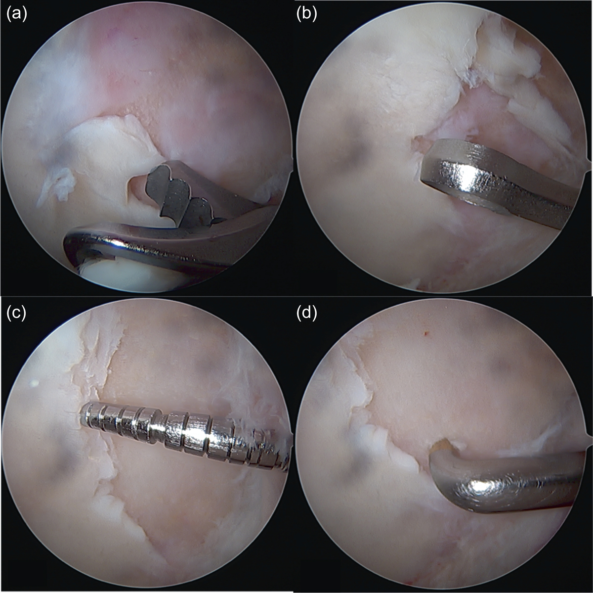
Treatment of the full-thickness focal chondral lesion: (a) Resection of the unstable chondral tissue is performed in the first instance with either shaver or tissue resection. (b) The unstable edges of the lesion are then resected with a ringed curette, leaving the edge as stable and vertical as possible; also the calcareous layer of the exposed subchondral bone is removed with the curettes. (c) After this procedure, it is recommended to palpate the edges of the lesion to confirm the stability of the remaining tissue as well as to measure the chondral defect in millimetric scale. (d) The microfractures are then made with an arthroscopic awl beginning at the vertex of the lesion. For this purpose, there are awls of different angulation to make the perforations as vertical as possible to the lesion.
Current usage metrics show cumulative count of Article Views (full-text article views including HTML views, PDF and ePub downloads, according to the available data) and Abstracts Views on Vision4Press platform.
Data correspond to usage on the plateform after 2015. The current usage metrics is available 48-96 hours after online publication and is updated daily on week days.
Initial download of the metrics may take a while.