Figure 1.
Download original image
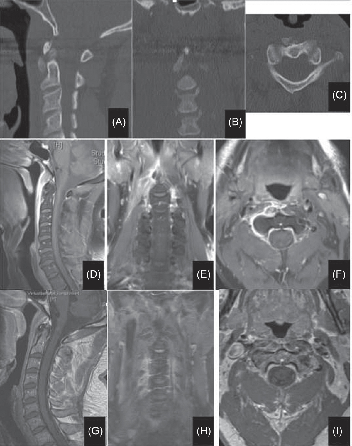
(A–C) CT scans showing calcifications at longus colli origin anterior to C1/2, measuring about 14 mm in the superior-inferior diameter and about 7 mm in the anteroposterior diameter. (D–F) Prevertebral swelling and minimal fluid collection extending from skull base caudally to C4, maximally at the middle of C2 (up to 7 mm). Musculus longus colli on the right side is edematous especially at its insertion at C1 and C2 vertebrae. No other sites of inflammation in the ENT area. The findings correspond to right-sided longus colli tendinitis. (G–I) A follow-up MRI with the same sequences shows the obvious regression of the soft tissue swelling as well as the contrast enhancement. Note: the MRIs were done using two different devices and the mostly corresponding cuts were selected for the comparison between that at presentation and at follow-up.
Current usage metrics show cumulative count of Article Views (full-text article views including HTML views, PDF and ePub downloads, according to the available data) and Abstracts Views on Vision4Press platform.
Data correspond to usage on the plateform after 2015. The current usage metrics is available 48-96 hours after online publication and is updated daily on week days.
Initial download of the metrics may take a while.