Figure 2
Download original image
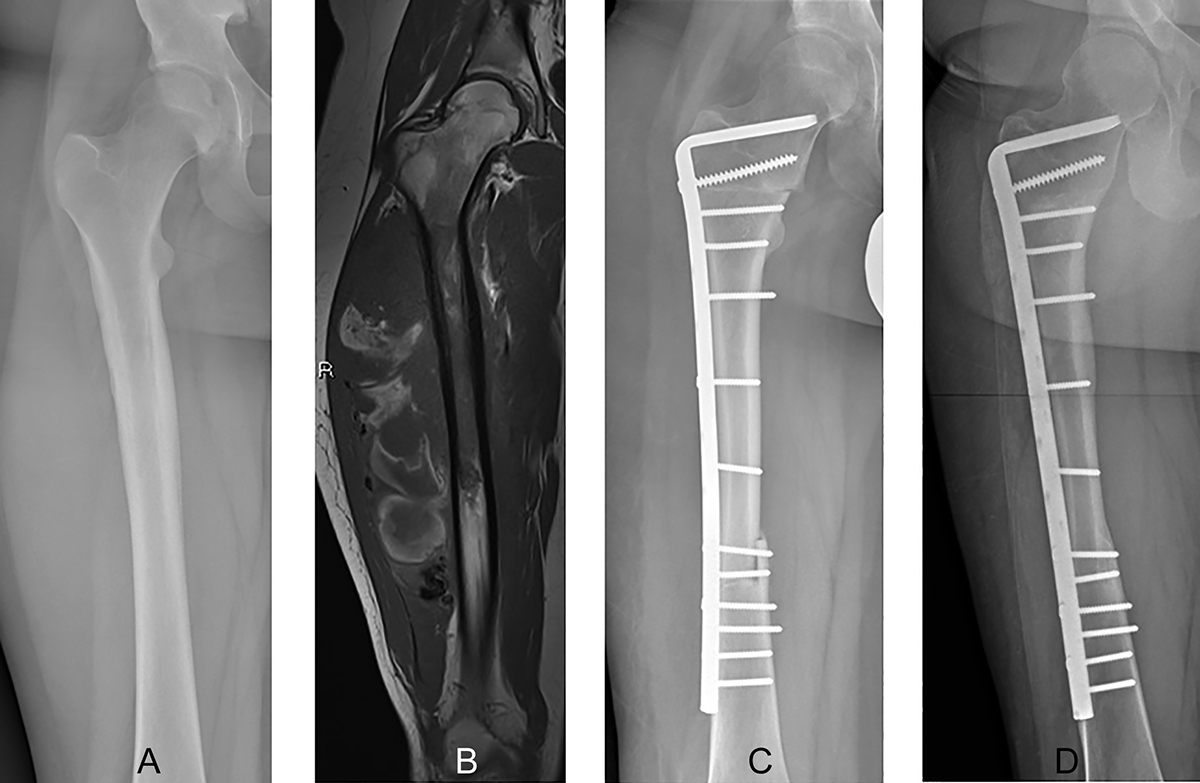
A 16-year-old boy with a diagnosis of Ewing sarcoma of the right femur. Preoperative anteroposterior radiographic view (A) showing a lytic lesion with periosteal reaction at the proximal diaphyseal region of the right femur. Coronal-T1 (B) magnetic resonance image shows tumor extension and a large extraosseous mass at the proximal diaphysis without involvement of the meta-epiphyseal region of the femur. Radiographs showing intercalary reconstruction of the femur using a massive bone allograft at the three-month (C) and 8-year (D) follow-up, showing consolidation and integration of the massive bone allograft. The massive bone allograft was able to be incorporated into the host bone despite the absence of the vascularized fibula graft as the resection was less than 15 cm.
Current usage metrics show cumulative count of Article Views (full-text article views including HTML views, PDF and ePub downloads, according to the available data) and Abstracts Views on Vision4Press platform.
Data correspond to usage on the plateform after 2015. The current usage metrics is available 48-96 hours after online publication and is updated daily on week days.
Initial download of the metrics may take a while.