Figure 3
Download original image
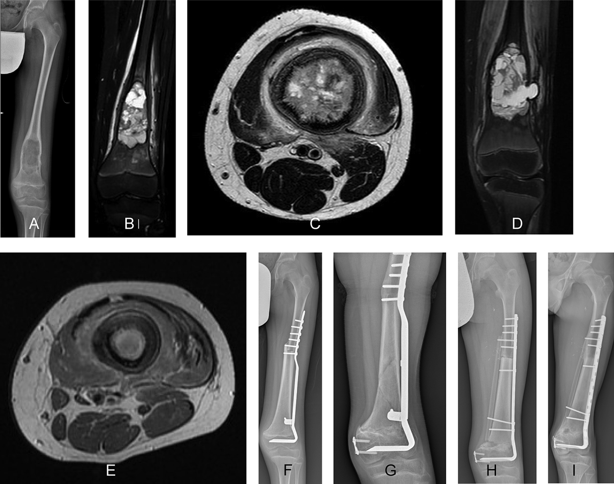
A 12-year-old boy, diagnosed with osteosarcoma of the left femur, was treated with intercalary resection and massive bone allograft reconstruction. (A) An anteroposterior radiographic view at presentation showing a lytic lesion located at the distal diaphyseal region of the femur, without involvement of the physis. The edema around the lesion seen in the anteroposterior T2 Short-TI Inversion Recovery magnetic resonance image (B) and the axial T2 magnetic resonance image (C) before preoperative chemotherapy disappeared in the anteroposterior T2 Short-TI Inversion Recovery magnetic resonance image (D) and the axial T2 magnetic resonance image (E) after preoperative chemotherapy. (F) Radiograph showing consolidation of the intercalary allograft at the 3-month follow-up. Radiograph showing allograft fracture 2 years after the surgery (G) treated using a new massive bone allograft in combination with a free vascularized fibular graft (H). (I) Radiographic view showing consolidation and integration of the allograft with the fibular graft 2 years after the surgery. The vascularized fibula graft could serve as an alternative for addressing fracture of intercalary massive bone allograft.
Current usage metrics show cumulative count of Article Views (full-text article views including HTML views, PDF and ePub downloads, according to the available data) and Abstracts Views on Vision4Press platform.
Data correspond to usage on the plateform after 2015. The current usage metrics is available 48-96 hours after online publication and is updated daily on week days.
Initial download of the metrics may take a while.