Figure 6
Download original image
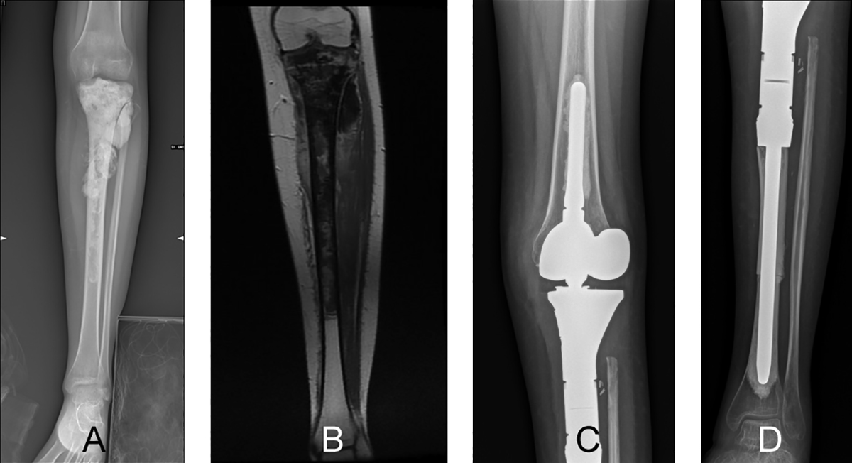
A 17-year-old girl diagnosed with osteosarcoma of the left proximal tibia who was treated by osteoarticular resection and modular prosthesis reconstruction. (A) Preoperative anteroposterior radiographic view showing a permeating sclerotic lesion of the proximal tibia. (B) A coronal-T1 magnetic resonance image showing the tumor located at the proximal tibia that extends to the diaphysis. Postoperative radiographs (C and D) show modular prosthesis reconstruction and intercalary massive bone allograft augmentation of the proximal tibia at the 12-month follow-up. The modular prosthesis was one of the treatments of choice in children at the end of their skeletal growth undergoing joint resection for bone sarcoma.
Current usage metrics show cumulative count of Article Views (full-text article views including HTML views, PDF and ePub downloads, according to the available data) and Abstracts Views on Vision4Press platform.
Data correspond to usage on the plateform after 2015. The current usage metrics is available 48-96 hours after online publication and is updated daily on week days.
Initial download of the metrics may take a while.