Figure 7
Download original image
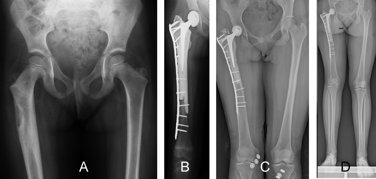
A 4-year-old girl diagnosed with Ewing sarcoma of the right proximal femur treated with osteoarticular resection and allograft–prosthesis composite reconstruction. (A) A preoperative radiograph showing an osteolytic lesion present in the subtrochanteric region of the proximal femur. (B) A postoperative radiograph shows reconstruction, using an allograft–prosthesis composite with a short stem and plate fixation together with reconstruction of the hip with a unipolar hemiarthroplasty with femoral ceramic head. Anteroposterior radiographic view (C) after 16 years of follow-up showing consolidation of the allograft, no loosening of the femoral prosthesis stem, growth of the remaining distal femur, and fracture of the greater trochanter of the allograft, and panoramic view (D) showing a discrepancy of 1.80 cm in limb length. Allograft–prosthesis composites may be an alternative to modular prosthesis in children with bone sarcoma of the proximal femur who have a small size of bone.
Current usage metrics show cumulative count of Article Views (full-text article views including HTML views, PDF and ePub downloads, according to the available data) and Abstracts Views on Vision4Press platform.
Data correspond to usage on the plateform after 2015. The current usage metrics is available 48-96 hours after online publication and is updated daily on week days.
Initial download of the metrics may take a while.