Figure 8
Download original image
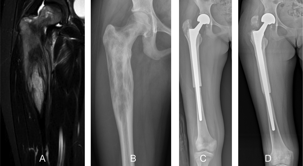
A 13-year-old girl, diagnosed with osteosarcoma, was treated with osteoarticular resection of the right proximal femur followed by reconstruction using an allograft–prosthesis composite. A coronal-T1 (A) magnetic resonance image shows a tumor extension to the proximal meta-diaphyseal region of the right femur. A preoperative anteroposterior radiographic view (B) showing an osteolytic lesion of the proximal femur. (C) Radiograph at one month postoperatively, showing the reconstruction using a press-fit femoral stem and reconstruction of the hip with bipolar hemiarthroplasty. (D) Radiograph at the 1-year follow-up showing fracture of the greater trochanteric region. Allograft–prosthesis composites may help preserve bone stock for future revision surgeries in children with bone sarcoma of the proximal femur.
Current usage metrics show cumulative count of Article Views (full-text article views including HTML views, PDF and ePub downloads, according to the available data) and Abstracts Views on Vision4Press platform.
Data correspond to usage on the plateform after 2015. The current usage metrics is available 48-96 hours after online publication and is updated daily on week days.
Initial download of the metrics may take a while.