Figure 1

Download original image
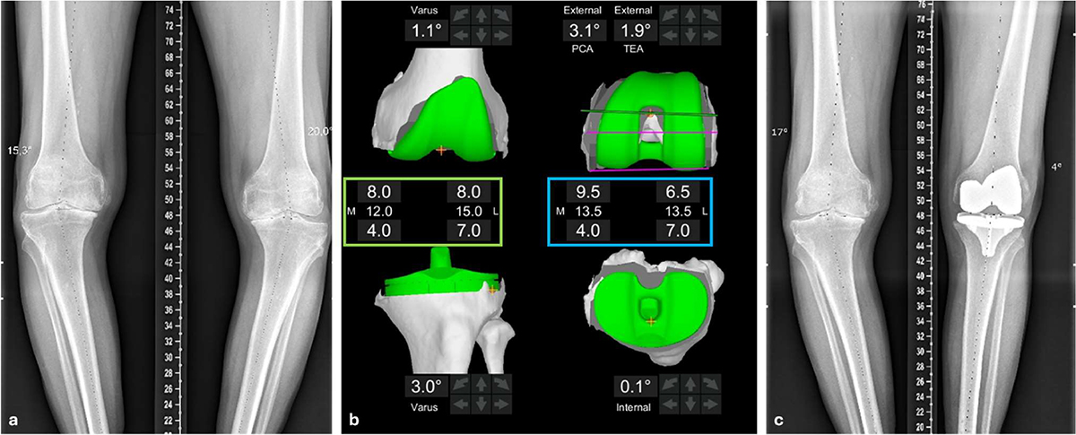
Management of a patient with a bilateral knee osteoarthritis with large deformity. a: preoperative long leg standing X-rays showing a varus deformation of 20°. (b) Intra-operative Mako settings. Coronal femoral component positioning is set to restore the native joint surface. Coronal tibial component positioning is set to obtain an equal medial and lateral extension gap. The green box represents the value of the distal femoral cuts (values at the top of the box), and the proximal tibia cuts (values at the bottom of the box). The middle values represent the total cut thickness. For this case, femoral varus is set at 1.1° and tibial varus is set at 3° to achieve this goal. In the axial plane, femoral component is positioned to align with the trochlear groove while having an equal medial and lateral flexion gap. The blue box represents the thickness of the posterior femoral cuts (values at the top of the box) and the proximal tibial cuts (values at the bottom of the box). The middle values represent the total cut thickness. For this case, femoral component rotation is set at 3.1° relative to the PCA (Posterior Condyle Axis) or 1.9° relative to the sTEA (surgical Trans-Epicondylar Axis). (c) One-year postoperative long-leg standing X-rays showing a global varus value of 4°.
Current usage metrics show cumulative count of Article Views (full-text article views including HTML views, PDF and ePub downloads, according to the available data) and Abstracts Views on Vision4Press platform.
Data correspond to usage on the plateform after 2015. The current usage metrics is available 48-96 hours after online publication and is updated daily on week days.
Initial download of the metrics may take a while.


