Figure 1
Download original image
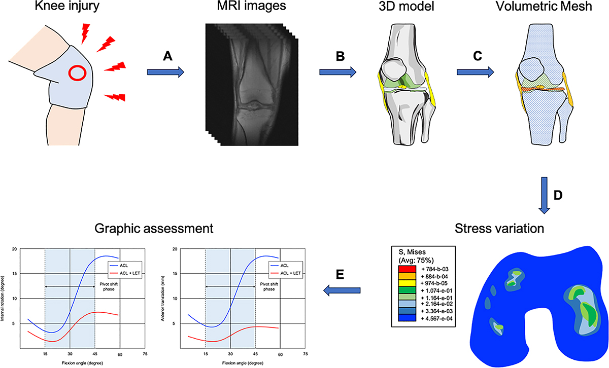
A graphical overview of the finite element analysis of the knee joint. A) A magnetic resonance imaging (MRI) is performed after knee injury. B) A three-dimensional (3D) model is created using computed tomography (CT) or/and MRI of the knee joint. C) The volumetric geometries of the knee joint are discretized for analysis. D) Contact stress behaviour of the femoral condyles after anterior cruciate ligament rupture. E) The profile of pivot shift phase after ACLr with and without LET.
Current usage metrics show cumulative count of Article Views (full-text article views including HTML views, PDF and ePub downloads, according to the available data) and Abstracts Views on Vision4Press platform.
Data correspond to usage on the plateform after 2015. The current usage metrics is available 48-96 hours after online publication and is updated daily on week days.
Initial download of the metrics may take a while.