Figure 2
Download original image
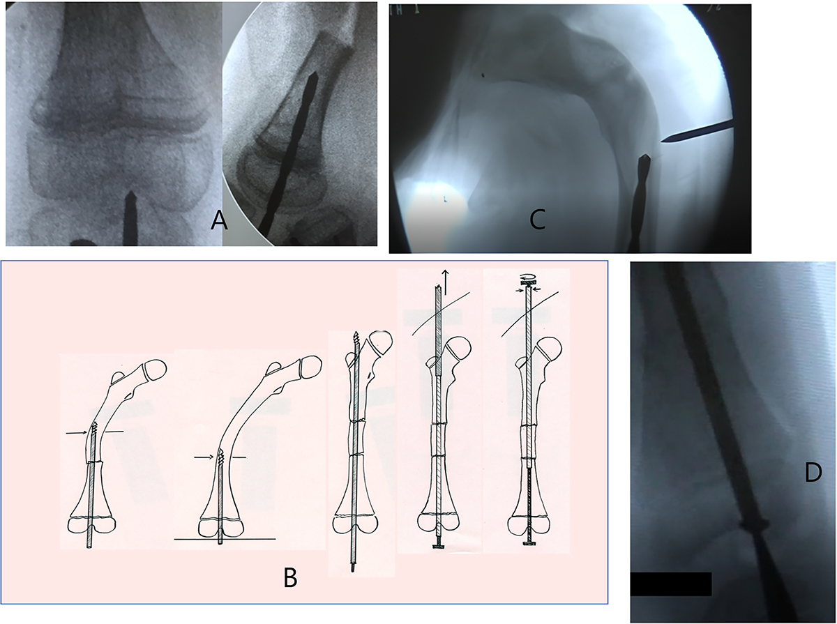
This figure illustrates the surgical technique. A: Manual drilling is performed perpendicular to the joint line in both anterior (left) and lateral (right) views. B: During drilling, it is crucial to remain perpendicular to the distal femoral joint line, performing osteotomies as needed. These can be done either via percutaneous osteoclasis or through a lateral open approach (this figure was created by the authors to be used in this article). C: The exit point for the drill bit is at the femoral neck, allowing for valgus fixation to prevent coxa vara deformity and the risk of lateral nail exit or femoral neck fracture. To facilitate this, a subtrochanteric osteotomy may be performed. D: The male component is introduced percutaneously through the knee and impacted into the distal femoral epiphysis.
Current usage metrics show cumulative count of Article Views (full-text article views including HTML views, PDF and ePub downloads, according to the available data) and Abstracts Views on Vision4Press platform.
Data correspond to usage on the plateform after 2015. The current usage metrics is available 48-96 hours after online publication and is updated daily on week days.
Initial download of the metrics may take a while.