Figure 6
Download original image
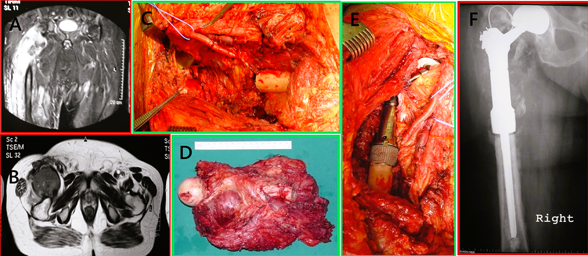
Preoperative imaging, intraoperative steps, and postoperative outcome of proximal femoral replacement in a 69-year-old male with recurrent high-grade leiomyosarcoma of the right thigh. (A) Coronal MRI demonstrating a recurrent, aggressive soft tissue mass infiltrating the proximal right femur with cortical destruction and intramedullary extension. (B) Axial MRI showing the extent of soft tissue involvement and medullary infiltration. (C) Intraoperative photograph following wide en bloc resection of the proximal femur. (D) Gross specimen showing the resected proximal femur with the attached high-grade leiomyosarcoma mass. (E) Intraoperative image during implantation of a modular Stanmore METS® proximal femoral megaprosthesis. (F) Immediate postoperative anteroposterior radiograph of the right hip demonstrating final reconstruction with the Stanmore METS® implant in situ.
Current usage metrics show cumulative count of Article Views (full-text article views including HTML views, PDF and ePub downloads, according to the available data) and Abstracts Views on Vision4Press platform.
Data correspond to usage on the plateform after 2015. The current usage metrics is available 48-96 hours after online publication and is updated daily on week days.
Initial download of the metrics may take a while.