Figure 1
Download original image
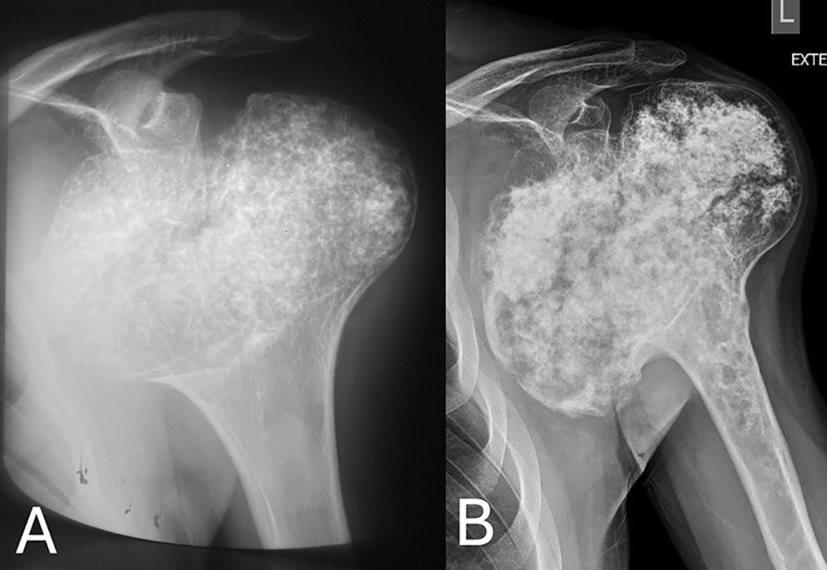
(A) Anteroposterior plain radiograph of left shoulder at the time of initial diagnosis (age 17), showing a bulky bilobed symmetric osteocartilagenous overgrowth of the proximal humeral epiphysis with stippled calcifications, cortical thinning, and a sharp zone of transition towards the metaphysis. (B) Anteroposterior plain radiograph of the left shoulder revealing the bulky, bilobed, incongruent osteocartilagenous overgrowth of the proximal humeral epiphysis. The overgrowth was asymmetric with exuberant enlargement of the medial component, which presented osteolytic areas and areas of cortical breach. Rings and arcs, calcifications, and osteolytic foci were evident in the proximal metaphysis.
Current usage metrics show cumulative count of Article Views (full-text article views including HTML views, PDF and ePub downloads, according to the available data) and Abstracts Views on Vision4Press platform.
Data correspond to usage on the plateform after 2015. The current usage metrics is available 48-96 hours after online publication and is updated daily on week days.
Initial download of the metrics may take a while.