Figure 2
Download original image
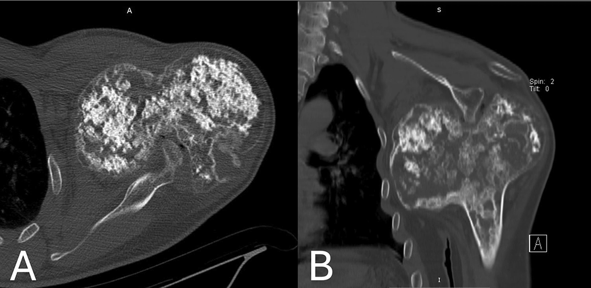
A & B. Computed tomography scan images showing the presence of extensive cartilaginous calcifications, cortical remodeling, and an osteolytic area with cortical disruption at the posterior aspect of the epiphyseal overgrowth. The medial epiphyseal overgrowth extended to the maxillary recess, whereas the glenoid process was seen to indent to the posterior aspect of the epiphysis.
Current usage metrics show cumulative count of Article Views (full-text article views including HTML views, PDF and ePub downloads, according to the available data) and Abstracts Views on Vision4Press platform.
Data correspond to usage on the plateform after 2015. The current usage metrics is available 48-96 hours after online publication and is updated daily on week days.
Initial download of the metrics may take a while.