


Figure 2
Download original image
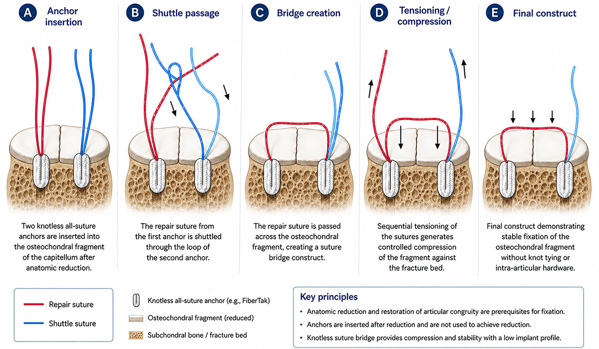
Schematic illustration of the reduction-first knotless fixation technique. (A) Insertion of two knotless all-suture anchors after anatomic reduction of the osteochondral fragment. (B) Shuttle passage of the repair sutures through the corresponding anchor loops. (C) Creation of the suture bridge construct across the fragment. (D) Sequential tensioning generates controlled compression across the fracture bed. (E) Final low-profile construct providing stable fixation without intra-articular metal hardware or knot stacks.
Current usage metrics show cumulative count of Article Views (full-text article views including HTML views, PDF and ePub downloads, according to the available data) and Abstracts Views on Vision4Press platform.
Data correspond to usage on the plateform after 2015. The current usage metrics is available 48-96 hours after online publication and is updated daily on week days.
Initial download of the metrics may take a while.