Figure 5.

Download original image
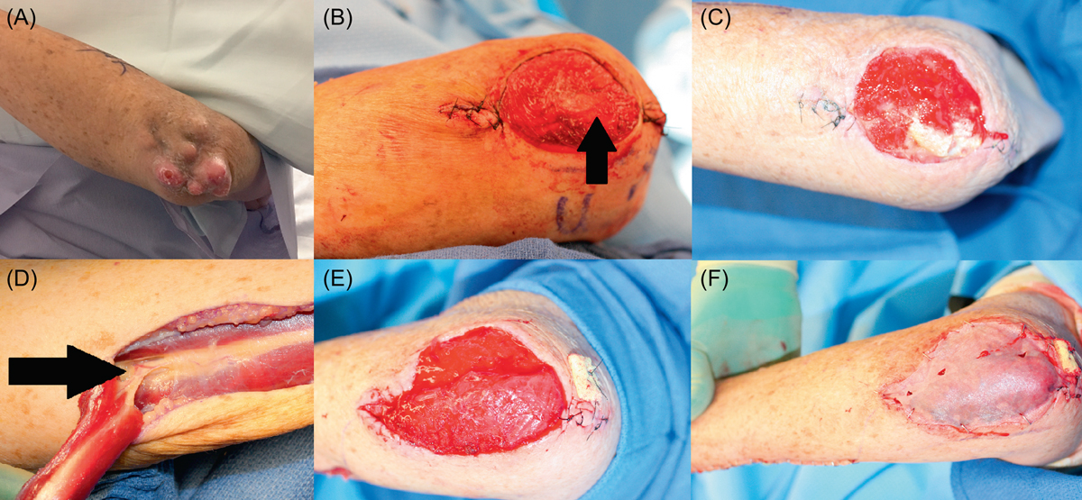
Seventy-one-year-old male with a myxoid sarcoma of the left elbow area. (A) Painless mass developing over the course of a few years with local progression and the development of satellite lesions. (B) Resection of the tumor including the periosteum (arrow) of the ulnar bone. As the examination of the margins was pending, this wound was temporarily protected by the subsequent application of a VAC-system. (C) One week after primary resection: the defect remains non-infected and prepared for reconstructive surgery. A muscle flap was necessary for sufficient coverage of the exposed bone and to provide well-vascularized coverage in case of adjuvant radiotherapy as part of the treatment protocol or in case of a recurrence. A skin graft would not have offered adequate durability attributes and would have been unlikely to heal over the periosteum-denuded bone. (D) Dissection of the flexor carpi ulnaris muscle flap: major vascular pedicle entering the deep surface of the muscle 6 cm distal to the elbow (arrow) with the visible delicate anastomoses forming the cubital vascular rete. (E) Defect coverage with the muscle flap. The distal tendon was embedded underneath the proximal wound edge and fixed percutaneously to a gauze bolster to ensure healing without flap dislodgment. (F) Split thickness skin graft covering the muscle flap surface.
Current usage metrics show cumulative count of Article Views (full-text article views including HTML views, PDF and ePub downloads, according to the available data) and Abstracts Views on Vision4Press platform.
Data correspond to usage on the plateform after 2015. The current usage metrics is available 48-96 hours after online publication and is updated daily on week days.
Initial download of the metrics may take a while.


