Figure 1.
Download original image
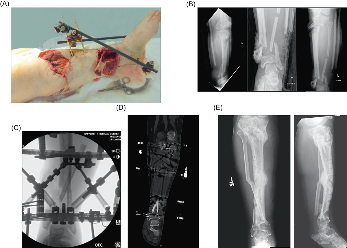
Case example of a 49-year-old male polytrauma who sustained a Gustilo-Anderson grade IIIB open left proximal and distal tibial fractures following airplane crash. (A) Clinical images of large anteromedial wounds over the proximal and distal thirds of the leg with segmental bone loss distally, (B) anteroposterior and lateral injury radiographs demonstrating the aforementioned injuries with associated fibular fracture, (C) initial post-operative fluoroscopic images with Taylor Spatial Frame (TSF) in place, (D) coronal CT renderings of the distal tibial fracture at the time of nonunion (seven months following injury) with Taylor Spatial Frame in place, and (E) final film taken one year following RIA at the time of union and TSF removal.
Current usage metrics show cumulative count of Article Views (full-text article views including HTML views, PDF and ePub downloads, according to the available data) and Abstracts Views on Vision4Press platform.
Data correspond to usage on the plateform after 2015. The current usage metrics is available 48-96 hours after online publication and is updated daily on week days.
Initial download of the metrics may take a while.