Figure 4
Download original image
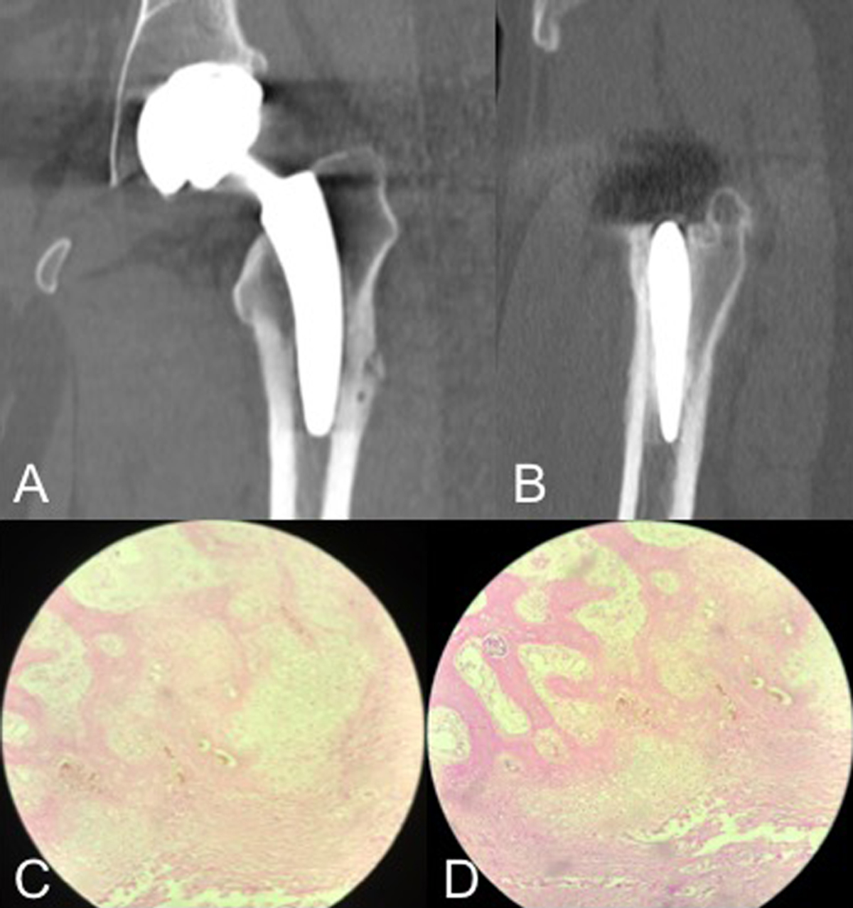
A and B. Coronal and sagittal computed tomography slices obtained at the time of bone biopsy, taken from the lateral femoral cortical at 9 months of follow-up depicting no evidence of stem loosening or sinking. C and D. 10x and 40x haematoxylin and eosin staining histological sections taken from a bone biopsy taken at the lateral femoral cortical of the patient's left hip at 9 months of follow-up. Pathology analysis reveals disorganized trabecular bone and cartilaginous tissue with peri-haversian bone formation areas and abundant capillaries (angiogenesis), as well as increased subperiosteal osteoblastic activity.
Current usage metrics show cumulative count of Article Views (full-text article views including HTML views, PDF and ePub downloads, according to the available data) and Abstracts Views on Vision4Press platform.
Data correspond to usage on the plateform after 2015. The current usage metrics is available 48-96 hours after online publication and is updated daily on week days.
Initial download of the metrics may take a while.