Figure 3
Download original image
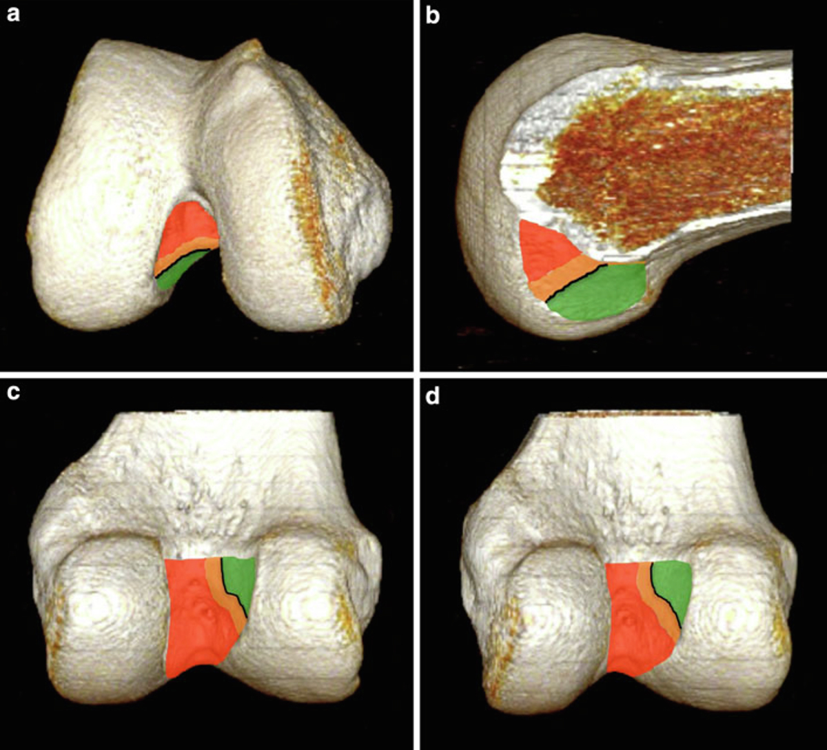
3-D CT scan reconstruction of the right distal femur illustrating the femoral tunnel classification system for the anterior cruciate ligament (ACL). a. Distal view. b. Medial view. c. Posterior view. d. Posteromedial view. Type I tunnels (appropriately positioned or anatomical) are entirely contained within the green zone, located posterior and inferior to the lateral intercondylar ridge (black line). Type II tunnels (slightly malpositioned) partially overlap the lateral intercondylar ridge and extend into the orange zone. Type III tunnels (severely malpositioned or non-anatomical) are positioned completely anterior and superior to the lateral intercondylar ridge, extending into the red zone.
Current usage metrics show cumulative count of Article Views (full-text article views including HTML views, PDF and ePub downloads, according to the available data) and Abstracts Views on Vision4Press platform.
Data correspond to usage on the plateform after 2015. The current usage metrics is available 48-96 hours after online publication and is updated daily on week days.
Initial download of the metrics may take a while.