


| Issue |
SICOT-J
Volume 12, 2026
|
|
|---|---|---|
| Article Number | 41 | |
| Number of page(s) | 6 | |
| Section | Hip | |
| DOI | https://doi.org/10.1051/sicotj/2026038 | |
| Published online | 03 July 2026 | |
Case Report
Atypical femoral fracture: The periprosthetic variant about two cases without bisphosphonate use
Diaconesses Croix Saint-Simon Hospital Group, 125 rue d’Avron, 75020 Paris, France
* Corresponding author: This email address is being protected from spambots. You need JavaScript enabled to view it.
Received:
11
February
2026
Accepted:
6
May
2026
Abstract
Atypical femoral fractures are rare. They are well described in patients with prolonged bisphosphonate use. The periprosthetic form without bisphosphonate intake appears to be exceptional and may lead to mechanical complications (nonunion, secondary displacement) if the difficulties of bone healing are not anticipated. We report here two cases of atypical femoral fractures without bisphosphonate use and the resulting implications for surgical fixation.
Key words: Periprosthetic fracture / Atypical femoral fracture / AFF
© The Authors, published by EDP Sciences, 2026
 This is an Open Access article distributed under the terms of the Creative Commons Attribution License (https://creativecommons.org/licenses/by/4.0), which permits unrestricted use, distribution, and reproduction in any medium, provided the original work is properly cited.
Introduction
Although rare, atypical femoral fractures (AFFs) pose a real challenge for both diagnosis and medical-surgical management. The American Society for Bone and Mineral Research (ASBMR) has long described major and minor criteria to aid in the diagnosis of such fractures (Table 1).
(4 major criteria needed) Diagnostic criteria for atypical femoral fractures.
The main causes of AFFs reported in the literature are the use of osteoclast inhibitors, particularly bisphosphonates and denosumab, usually after prolonged use, which alters bone remodeling.
The pathogenesis of AFFs remains poorly understood [1]. However, the literature consistently highlights a strong association with long-term bisphosphonate use.
We present two cases of female patients who met all diagnostic criteria for AFFs, but whose medical history did not include any causal medication. Both had unexplained hip prosthesis pain for several weeks. These cases raise questions about the pathophysiology of atypical femoral fractures and confirm the current recommendations regarding the need for rigid fixation strategies.
Case presentations
Case 1
An 86-year-old female was hospitalized for advanced bicompartmental knee osteoarthritis unresponsive to conservative treatment. She was 1.48 m tall and weighed 85 kg (BMI: 38 kg/m2). A total left knee replacement was indicated. Her medical history included:
Restless legs syndrome.
Right total hip arthroplasty for osteoarthritis (2022).
Laminectomy L4–L5 (2022).
Left femoral shaft fracture, treated by intramedullary nailing and later bone grafting (2020).
Her medications included pramipexole, gabapentin, and duloxetine.
Preoperatively, she reported contralateral thigh pain, which was initially managed conservatively (rest and analgesics).
A left total knee arthroplasty (Attune®, Johnson & Johnson®) was performed with intraoperative navigation and kinematic alignment (Varus 12°–4°) (Figure 1). Postoperatively, she was mobilized but continued to experience contralateral thigh pain. She was discharged to a rehabilitation facility.
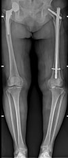 |
Figure 1 Incomplete atypical femoral fracture. |
Shortly thereafter, the patient reported sudden functional impairment while sitting, without trauma. On readmission, radiographs revealed a right periprosthetic femoral fracture classified as AO/OTA 32A3, Vancouver C (Figure 2).
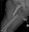 |
Figure 2 Complete atypical femoral fracture. |
Pre-injury panoramic radiographs showed cortical thickening at the tip of the stem, followed by a stress fracture.
The patient underwent open reduction and internal fixation via a lateral subvastus approach using a Zimmer® NCB Periprosthetic plate with a 15 cm distal lever arm, 10-cortex screw fixation (distal plate screw density 0.83), and high-density proximal locking screws (Figure 3). Working length was nearly 3 cm.
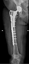 |
Figure 3 Internal fixation of the femoral fracture. |
She developed a postoperative pulmonary embolism, managed with curative anticoagulation (tinzaparin 14,000 IU anti-Xa/day, then apixaban 5 mg BID).
At 45 days post-op, full weight-bearing was authorized.
However, at day 82, she reported new groin and thigh pain without trauma. Imaging showed secondary displacement and plate failure (Figure 4).
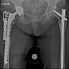 |
Figure 4 Iterative femoral Fracture and material breakage. |
She underwent revision surgery with a longer lateral plate, an anterior neutralization plate, and a posterior bone allograft (tibial strut) (Figure 5)
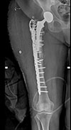 |
Figure 5 Iterative internal fixation enhanced with allograft. |
Histopathology from the fracture site revealed hypertrophic bone repair tissue with fibrous and cartilaginous components; no tumor or inflammation was found. Bacteriological cultures (3/3) were sterile.
Postoperative protocol included toe-touch weight-bearing for 6 weeks. Bone healing was achieved at 6 months (Figure 6), and the patient resumed daily activities pain-free at the last follow-up (15 months).
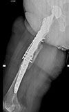 |
Figure 6 Posterior union on the lateral X-ray. |
Case 2
Another 86-year-old female with a history of:
Marfan syndrome.
Breast cancer in remission.
Left total knee arthroplasty.
Bilateral total hip replacements for osteoarthritis (2010).
She measured 1.60 m, weighed 64 kg (BMI: 25 kg/m2), and her medications included prednisone, clopidogrel, and candesartan.
She consulted for persistent pain over her left hip prosthesis for 1 year. Radiographs were reported as normal (Figure 7), and a bone scan and articular injection were scheduled.
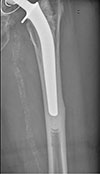 |
Figure 7 Incomplete atypical femoral fracture. |
Seventeen days later, she returned with total functional impairment and visible thigh deformity without any traumatic event.
X-rays revealed a transverse left femoral fracture (AO/OTA 32A3) at the stem tip, Vancouver C (Figure 8).
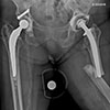 |
Figure 8 Complete atypical femoral fracture. |
Given her clinical history, an allograft was planned upfront. Surgery was performed 2 days later: Open reduction and dual-plate fixation were performed with a Zimmer® NCB periprosthetic plate laterally (~20 cm distal lever arm; distal plate screw density 0.75), and a second orthogonal Synthes® plate. Posterior and medial strut allografts were added and secured with screws (Figure 9).
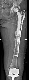 |
Figure 9 Primary internal fixation enhanced with allograft. |
She was discharged to a geriatric perioperative unit without complications. Toe-touch weight-bearing was permitted. At the 14-week follow-up, the patient was pain-free and allowed full weight-bearing. Radiographs showed early callus formation (Figure 10).
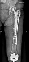 |
Figure 10 Union of the atypical femoral fracture. |
Discussion
Rare entity
Atypical femoral fractures are rare and poorly understood. More frequently reported in Asian populations, they are commonly associated with bisphosphonate therapy [2].
Periprosthetic AFFs have been described but are almost always linked to bisphosphonate use, as in a recent retrospective study of all periprosthetic fractures in a single center [3].
To our knowledge, this is the first case series of periprosthetic AFFs without any causative drug history.
Notably, in both cases, radiographs prior to the fracture were available.
Pathophysiology
Bisphosphonates may compromise the femoral nutrient artery endothelium, generating a focal anterolateral stress concentration that can lead to fatigue fractures.
Analogous to the mechanics of felling a tree, where loading follows gravity lines, prosthetic stem tips may create a similar stress riser, particularly in curved femurs [4].
These fractures are low-energy, bending-type injuries, with no rotational component, explaining their classic non-comminuted morphology.
These two cases had a potential confounding factor. The first patient likely increased the load on the right lower limb due to the recent implantation of a contralateral knee prosthesis, and the second patient had Marfan syndrome and took corticotherapy, which can increase the risk of fracture.
Management
Surgical treatment
AFFs are mechanically unstable and more prone to complications than typical femoral fractures [5], including nonunion and secondary displacement.
It is now established that fixation must be as rigid as possible (enhance the lever arm, diminish the working length, augment with an allograft). In the first case, the fixation used was a standard construct with 10 cortices on each side and a high screw density (83%), but without bone grafting. The rigidity of the construct was too low, and the fracture did not have enough time to heal before failing.
In native bone, intramedullary nails with multiple interlocking screws are preferred. For periprosthetic fractures, long plates with high-density screw fixation are required.
Adding an orthogonal second plate is often recommended for increased stability.
Allografts may enhance both mechanical strength (acting as a plate, allowing the sharing of load) and biological healing.
While replacing the stem with a long revision implant is a theoretical option, the risks of femoral osteotomy and implant-related complications make this a complex decision in periprosthetic cases.
Medical treatment
While bisphosphonates are implicated in AFFs, their role in preventing fragility fractures remains well-supported in the absence of contraindications.
Teriparatide has shown promise in enhancing the healing of AFFs but needs further high-quality studies.
Geriatric co-management improves postoperative outcomes in this frail population.
Contralateral and secondary prevention
Painful external cortical thickening, particularly at the stem tip, warrants consideration of preventive fixation.
A scoring system has been proposed to guide such decisions (indication if score ≥ 8) [6].
Conclusion
This case series highlights a unique presentation of periprosthetic atypical femoral fractures without prior bisphosphonate use.
The recommended surgical strategy is rigid internal fixation, recognizing the typically slow healing process.
Painful prosthetic implants with external cortical thickening should prompt evaluation for preventive osteosynthesis.
Funding
This research did not receive any specific funding
Conflicts of interest
Author 1: Consultant for Orthofix.
Author 2: None.
Author 3: Consultant for Depuy, Amplitude, royalties from Amplitude.
Author 4: Consultant for Depuy, Corin.
Author 5: None.
Data availability statement
All the data are available.
Author contribution statement
GA : Investigation, writing, original draft preparation.
TA: Editing and review.
SM: Writing and review.
YK: Editing and translation.
PL: Conceptualization, review and surgery.
Ethics approval
All procedures performed in studies involving human participants were in accordance with the ethical standards of the national research committee and with the 1964 Helsinki Declaration and its later amendments or comparable ethical standards. The study was approved by the local ethics committee and registered on ClinicalTrials.gov: CNIL MR004 2229500. The procedures used in this study adhere to the tenets of the Declaration of Helsinki. All patients gave informed consent to participate.
Informed consent
The 2 patients were informed and agreed to this work
References
- Tile L, Cheung AM (2020) Atypical femur fractures: current understanding and approach to management. Ther Adv Musculoskelet Dis.;Volume 12, 1–9 [Google Scholar]
- Gani L, Anthony N, Dacay L, Tan P, Chong LR, King TF (2021) Incidence of atypical femoral fracture and its mortality in a single center in Singapore. JBMR Plus 5, 1–9 [Google Scholar]
- Mondanelli N, Facchini A, Troiano E, Muratori F, Bottai V, Giannotti S (2021) Periprosthetic atypical femoral fractures exist: a retrospective study at a single Institution. Prevalence on 115 periprosthetic femoral fractures around a primary hip stem. J Arthroplasty 36, 2189–2196 [Google Scholar]
- W-T Cho, J-H Koh, S Sakong, J-T Kim (2025) Atypical femoral fractures: an update. J Muskuloskelet Trauma 38, 41–52. [Google Scholar]
- Baba T, Uchino M, Ochi H, Ikuta T, Saita Y, Hagino H, et al. (2021) Atypical periprosthetic femoral fractures after arthroplasty for fracture are at high risk of complications. Sci Rep https://doi.org/10.1038/s41598-021-93574-1 [Google Scholar]
- Koh A, Guerado E, Giannoudis PV (2017) Atypical femoral fractures related to bisphosphonate treatment: issues and controversies related to their surgical management. Bone Joint J 99, 295–302. [Google Scholar]
Cite this article as: Auberger G, Aubert T, Kerroumi Y, Leclerc P & Marmor S (2026) Atypical femoral fracture: The periprosthetic variant about two cases without bisphosphonate use. SICOT-J 12, 41. https://doi.org/10.1051/sicotj/2026038.
All Tables
All Figures
|
Figure 1 Incomplete atypical femoral fracture. |
| In the text | |
|
Figure 2 Complete atypical femoral fracture. |
| In the text | |
|
Figure 3 Internal fixation of the femoral fracture. |
| In the text | |
|
Figure 4 Iterative femoral Fracture and material breakage. |
| In the text | |
|
Figure 5 Iterative internal fixation enhanced with allograft. |
| In the text | |
|
Figure 6 Posterior union on the lateral X-ray. |
| In the text | |
|
Figure 7 Incomplete atypical femoral fracture. |
| In the text | |
|
Figure 8 Complete atypical femoral fracture. |
| In the text | |
|
Figure 9 Primary internal fixation enhanced with allograft. |
| In the text | |
|
Figure 10 Union of the atypical femoral fracture. |
| In the text | |
Current usage metrics show cumulative count of Article Views (full-text article views including HTML views, PDF and ePub downloads, according to the available data) and Abstracts Views on Vision4Press platform.
Data correspond to usage on the plateform after 2015. The current usage metrics is available 48-96 hours after online publication and is updated daily on week days.
Initial download of the metrics may take a while.