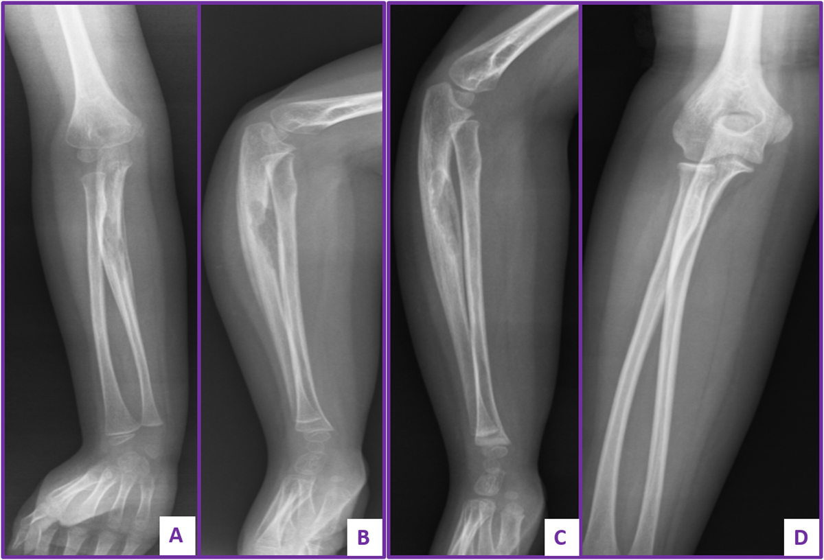
Figure 4
Download original image
Plain radiograph of forearm of a 5-year-old girl diagnosed with LCH showing large osteolytic lesion of the ulna managed conservatively. (A and B) Anteroposterior and lateral views showing the large sized osteolytic lesion of the ulna. (C) Follow-up after two years showing partial remodeling. (D) Follow-up radiograph showing complete remodeling.
Current usage metrics show cumulative count of Article Views (full-text article views including HTML views, PDF and ePub downloads, according to the available data) and Abstracts Views on Vision4Press platform.
Data correspond to usage on the plateform after 2015. The current usage metrics is available 48-96 hours after online publication and is updated daily on week days.
Initial download of the metrics may take a while.